Alternate header for print version
Contributors
Help
Submit
Search
menu
Data sets
Videos
Latest data
Center for Research in Biological Systems
Basic Science Building, Room 1000
University of California, San Diego
9500 Gilman Drive
La Jolla, CA 92093-0608, USA
Voice
: (858) 534-0276
Fax
: (858) 534-7497
Email
: dorloff@ncmir.ucsd.edu
Search Results for
retinal outer nuclear layer
(2970 results)
CIL:6517
NCBI Organism Classification
Danio rerio
Biological Process
retina layer formation
Cellular Component
none specified
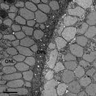
This study of the zebrafish eye is a series of four images taken at increasing magnifications. The first image is the SEM block face of the zebrafish eye. This is followed by this image, the second ...
CIL:6518
NCBI Organism Classification
Danio rerio
Biological Process
retina layer formation
Cellular Component
synapse
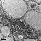
This study of the zebrafish eye is a series of four images taken at increasing magnifications. The first image is the SEM block face of the zebrafish eye. This is followed by a second image that is ...
CIL:10914
NCBI Organism Classification
Macaca mulatta
Biological Process
neurotransmitter secretion
Cellular Component
synapse part
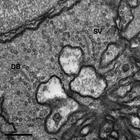
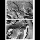
Freeze fracture images of synaptic junctions in the retina of Macaca mulatta. The electron micrograph in the top panel shows two invaginating synapses between cone and horizontal cells in the outer p...
CIL:11636
NCBI Organism Classification
Rattus sp.
Biological Process
epithelial cilium movement
Cellular Component
cilium
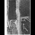
Figures 323 & 324 from Chapter 13 (Cilia and Flagella) of 'The Cell' by Don W. Fawcett M.D. The outer segments of the rods and cones of the vertebrate retina and many photoreceptors of invertebrates b...
CIL:38980
NCBI Organism Classification
Gallus gallus
Biological Process
eye development
Cellular Component
cell surface
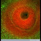
Confocal image of the developing eye of a chick embryo. The dark region in the centre is the where the surface layer has invaginated to form the lens vesicle. The tissue surrounding this space is stai...
CIL:49851
NCBI Organism Classification
Mus musculus
Biological Process
Intrinsic photosensitivity conferred by melanopsin photopigment
Cellular Component
cytoskeleton
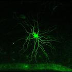
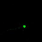
These color images show the three-dimensional morphology of an M5 melanopsin retinal ganglion cell from an <i>ex-vivo</i> isolated flat-mount mouse retina preparation. This retinal neuron was visually...
CIL:39022
NCBI Organism Classification
Danio rerio
Biological Process
optic nerve structural organization
Cellular Component
neuron projection
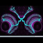
Confocal micrograph showing the connections of the visual system in a four-day-old zebrafish embryo. Staining of the neurons, glia and optic nerve illustrate the connections between the retina and the...
CIL:6519
NCBI Organism Classification
Danio rerio
Biological Process
synapse organization
Cellular Component
synapse
This study of the zebrafish eye is a series of four images taken at increasing magnifications. The first image is the SEM block face of the zebrafish eye. This is followed by a second image that is ...
CIL:57182
NCBI Organism Classification
Mus musculus
Biological Process
none specified
Cellular Component
cytoskeleton
The photopigment melanopsin confers photosensitivity upon a minority of retinal output neurons. These intrinsically photosensitive retinal ganglion cells (ipRGCs) are more diverse than once believed, ...
CIL:40380
NCBI Organism Classification
Solenostemon scutellarioides
Biological Process
plant stem organization
Cellular Component
cortex
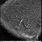
Scanning electron microscope image of cross-section through a Solenostemon scutellarioidesi (coleus) stem. The image shows the outer epidermal layer, cortex, vascular bundles in a ring, and a central ...
1
2
3
4
5
6
7
8
9
...
297
Next »
Results per page:
10
20
50
100

")