Alternate header for print version
Contributors
Help
Submit
Search
menu
Data sets
Videos
Latest data
Center for Research in Biological Systems
Basic Science Building, Room 1000
University of California, San Diego
9500 Gilman Drive
La Jolla, CA 92093-0608, USA
Voice
: (858) 534-0276
Fax
: (858) 534-7497
Email
: dorloff@ncmir.ucsd.edu
Search Results for
forebrain ventricle
(246 results)
CIL:39024
NCBI Organism Classification
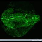
Danio rerio
Biological Process
gene expression
Cellular Component
PARD3
Confocal micrograph of a transgenic zebrafish embryo at 24 hours post-fertilization, showing expression of the fusion protein PARD3_GFP. PARD proteins, which were first identified in C. elegans, are e...
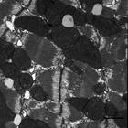
CIL:37212
NCBI Organism Classification
Rattus
Biological Process
regulation of cardiac muscle contraction
Cellular Component
mitochondrion
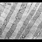
Transmission electron micrograph of longitudinal section of a a rat heart ventricle. Ordered arrays of myofibrils are interspersed with rows of mitochondria. Small, darkly staining glycogen granules a...
CIL:39781
NCBI Organism Classification
Mus musculus
Biological Process
muscle contraction
Cellular Component
I band
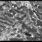
Transmission electron micrograph of ventricle tissue of a mouse cardiac muscle. This image shows the intercalated discs between the muscle fibers as well as many mitochondria and lipid droplets.
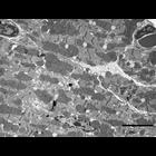
CIL:39772
NCBI Organism Classification
Mus musculus
Biological Process
cardiac muscle contraction
Cellular Component
mitochondrion
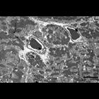
Transmission electron micrograph showing an over view of muscle tissue configuration from a mouse cardiac muscle ventricle. This image shows muscle to muscle connection and the abundance of mitochond...
CCDB:3603
Species
mouse
Organ
heart
Cell type
myocyte
System
cardiovascular
Structure
T-tubules, sarcoplasmic reticulum
MLP KO
CIL:39775
NCBI Organism Classification
Mus musculus
Biological Process
cardiac muscle fiber organization
Cellular Component
mitochondrion
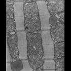
Transmission electron micrograph showing adjacent muscle fibers from the ventricle of a mouse cardiac muscle. This image shows the organization of muscle tissue, nucleus, cell walls and blood vessels...
CCDB:3611
Species
mouse
Organ
heart
Cell type
myocyte
System
cardiovascular
Structure
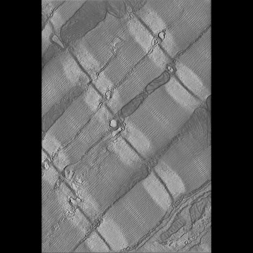
T-tubules, sarcoplasmic reticulum, mitochondrion
Experiment5
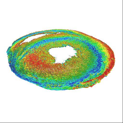
CCDB:9449
Species
Rat
Organ
heart
Cell type
none specified
System
cardiovascular
Structure
none specified
DT-MRI
CIL:39780
NCBI Organism Classification
Mus musculus
Biological Process
muscle contraction
Cellular Component
Z disc
Transmission electron micrograph of ventricle tissue of a mouse cardiac muscle. This image shows the various areas of a muscle fiber. The Z lines are the very dark borders that link sarcomeres. Th...
CIL:7567
NCBI Organism Classification
Myotis lucifugus
Biological Process
sarcomere organization
Cellular Component
mitochondrion
Cardiac muscle, particularly from the left ventricle, is rich in mitochondria since the heart requires an efficient continuous source of energy. In this highly magnified image, showing the length of t...
1
2
3
4
5
6
7
8
9
...
25
Next »
Results per page:
10
20
50
100