Alternate header for print version
Contributors
Help
Submit
Search
menu
Data sets
Videos
Latest data
Center for Research in Biological Systems
Basic Science Building, Room 1000
University of California, San Diego
9500 Gilman Drive
La Jolla, CA 92093-0608, USA
Voice
: (858) 534-0276
Fax
: (858) 534-7497
Email
: dorloff@ncmir.ucsd.edu
Search Results for
CeruleanFP
(5 results)
CIL:41631
NCBI Organism Classification
Mus musculus
Biological Process
none specified
Cellular Component
cytoplasm
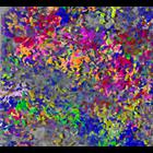
Confocal micrograph of NIH-3T3 cells co-transduced with 5 fluorescent proteins. The cells were marked by co-transduction with Lentiviral Gene Ontology (LeGO) vectors expressing Cerulean (cyan), EGFP ...
CIL:38652
NCBI Organism Classification
Homo sapiens
Biological Process
molecular organization
Cellular Component
focal adhesion
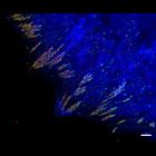
This image combines total internal reflection microscopy (TIRF) of mCerulean-actin (blue) with photoactivation localization microscopy (PALM) image of tdEos-vinculin (red) and Dronpa-paxillin (green)....
CIL:44153
NCBI Organism Classification
Mus musculus
Biological Process
clonal expansion of progenitor cells
Cellular Component
cytoplasm
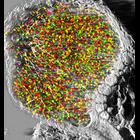
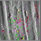
The picture represents a 3D-reconstruction (using Imaris software) of confocal/multiphoton (Leica SP5) images collected through the thickness (100 micrometers) of a live lymph node from a bone-marrow ...
CIL:44151
NCBI Organism Classification
Mus musculus
Biological Process
stem cell clonal tracking
Cellular Component
cytoplasm
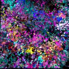
The image shows a 3D-rendering (Imaris software) of a live confluent culture of NIH-3T3 cells obtained using confocal microscopy. The cells were co-transduced with 5 fluorescent proteins and with Lent...
CIL:44152
NCBI Organism Classification
Mus musculus
Biological Process
clonal expansion of progenitor cells
Cellular Component
cytoplasm
The picture represents a 3D-reconstruction (using Imaris software) of confocal/multiphoton images collected through the thickness (100 micrometers) of the live skin from a bone-marrow transplanted mou...