Alternate header for print version
Contributors
Help
Submit
Search
menu
Data sets
Videos
Latest data
Center for Research in Biological Systems
Basic Science Building, Room 1000
University of California, San Diego
9500 Gilman Drive
La Jolla, CA 92093-0608, USA
Voice
: (858) 534-0276
Fax
: (858) 534-7497
Email
: dorloff@ncmir.ucsd.edu
Search Results for
protein glycosylation
(4976 results)
CIL:13553
NCBI Organism Classification
Homo sapiens
Biological Process
Golgi organization
Cellular Component
Golgi stack
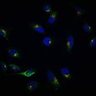
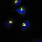
Helix Pomatia Lectin (HPL) (green) is redistributed out of the Golgi (Giantin) (red) in HeLa cells upon EGF stimulation for 4h. The Tn antigen refers to terminal α-linked N-acetyl galactosamine resid...
CIL:13544
NCBI Organism Classification
Homo sapiens
Biological Process
Golgi organization
Cellular Component
Golgi stack
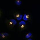
Helix Pomatia Lectin (HPL) (green) staining at the Golgi (Giantin) (red) in unstimulated HeLa cells. HPL binds various glycans but the Tn antigen in particular. The Tn antigen refers to terminal α-li...
CIL:13545
NCBI Organism Classification
Homo sapiens
Biological Process
Golgi organization
Cellular Component
Golgi stack
Anti-Tn staining (green) co-localizes with Helix Pomatia Lectin (HPL) (red) at the Golgi (Giantin) (gray) in unstimulated HeLa cells. The Tn antigen refers to terminal α-linked N-acetyl galactosamine...
CIL:13568
NCBI Organism Classification
Homo sapiens
Biological Process
retrograde vesicle-mediated transport, Golgi to ER
Cellular Component
Golgi-associated vesicle
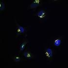
GalNac-T1 (green) is in dispersed punctate structures that stain positive for COP-I beta1 (red) after EGF treatment of HeLa cells for 4h. These data suggest that GalNAc-Ts are being redistributed from...
CIL:11382
NCBI Organism Classification
Mus musculus
Biological Process
post-translational protein modification
Cellular Component
Golgi apparatus
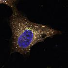
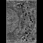
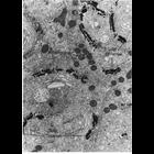
Figure 212 from Chapter 6 (Golgi Apparatus) of 'The Cell, 2nd Ed.' by Don W. Fawcett M.D. Secretory cells in Brunner's duodenal gland of the mouse have an extensive Golgi complex, with secretory granu...
CIL:11366
NCBI Organism Classification
Oniscus
Biological Process
post-translational protein modification
Cellular Component
Golgi apparatus
Figures 201 and 202 from Chapter 6 (Golgi Apparatus) of 'The Cell, 2nd Ed.' by Don W. Fawcett M.D. The Golgi apparatus of nurse cells from the testis of the woodlouse, Oniscus. Image by David Philli...
CIL:11372
NCBI Organism Classification
Chinchilla
Biological Process
post-translational protein modification
Cellular Component
Golgi apparatus
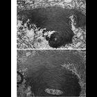
Figures 206 (upper) and 207 (lower) from Chapter 6 (Golgi Apparatus) of 'The Cell, 2nd Ed.' by Don W. Fawcett M.D. During development of the acrosome in the chinchilla spermatid, the transitional zon...
CIL:13547
NCBI Organism Classification
Homo sapiens
Biological Process
Golgi organization
Cellular Component
Golgi stack
GalNac-T1 staining (red) and Helix Pomatia Lectin (HPL) (green) colocalizes exclusively at the Golgi (Giantin) (gray) in unstimulated HeLa cells. The Tn antigen refers to terminal α-linked N-acetyl g...
CIL:11358
NCBI Organism Classification
Ovis aries
Biological Process
post-translational protein modification
Cellular Component
Golgi apparatus
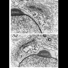
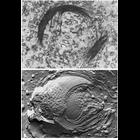
Figures 197 (upper) and 198 (lower) from Chapter 6 (Golgi Apparatus) of 'The Cell, 2nd Ed.' by Don W. Fawcett M.D. show electron microscopic views of the Golgi complex. Upper panel: A thin section pr...
CIL:11362
NCBI Organism Classification
Mus musculus
Biological Process
post-translational protein modification
Cellular Component
Golgi apparatus
Figure 199 from Chapter 6 (Golgi Apparatus) of 'The Cell, 2nd Ed.' by Don W. Fawcett M.D. The Golgi apparatus in epithelial cells from the mouse epididymis. The convex, or cis, side of the Golgi sho...
1
2
3
4
5
6
7
8
9
...
498
Next »
Results per page:
10
20
50
100