Alternate header for print version
Contributors
Help
Submit
Search
menu
Data sets
Videos
Latest data
Center for Research in Biological Systems
Basic Science Building, Room 1000
University of California, San Diego
9500 Gilman Drive
La Jolla, CA 92093-0608, USA
Voice
: (858) 534-0276
Fax
: (858) 534-7497
Email
: dorloff@ncmir.ucsd.edu
Search Results for
cell projection membrane
(35 results)
CIL:38957
NCBI Organism Classification
Homo sapiens
Biological Process
receiving nourishment
Cellular Component
zona pellucida
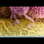
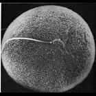
A colorized scanning electron micrograph of a human egg, which is the huge cell colored yellow at the bottom of this image. The follicle cells that surround it (top) send out long projections that pen...
CIL:39021
NCBI Organism Classification
none specified
Biological Process
cell-cell adhesion
Cellular Component
cell surface
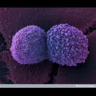
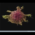
Colorized scanning electron micrograph showing two lung cancer cells. These cells were grown using cell culture techniques.
CIL:39049
NCBI Organism Classification
none specified
Biological Process
substrate-dependent cell migration, cell attachment to substrate
Cellular Component
cell surface
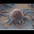
Colorized scanning electron micrograph of a lung cancer cell grown in culture.
CIL:11129
NCBI Organism Classification
Mus musculus
Biological Process
cell motility
Cellular Component
lamellipodium membrane
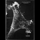
Scanning electron micrograph of a 3T3 cell growing in culture. Complexity and diversity of structure across the cell surface is apparent in the this image that s courtesy of Susan Brown. Figure 48 fr...
CIL:41920
NCBI Organism Classification
Mus musculus
Biological Process
synaptic connexion organization
Cellular Component
synaptic terminal
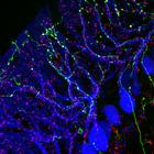
Confocal image of synaptic connexions in the mouse olivo-cerebellar system. Purkinje cells (immunofluorescence, blue), olivary axons (anterograde tracing, yellow) and synaptic terminals (immunofluore...
CIL:11094
NCBI Organism Classification
Phodopus
Biological Process
single fertilization
Cellular Component
microvillus membrane
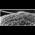
Scanning electron micrograph showing in vitro fertilization of a hamster oocyte. The sperm head, near the center of the field of view, is engulfed by the microvilli on the surface of the oocyte membr...
CIL:11096
NCBI Organism Classification
Phodopus
Biological Process
single fertilization
Cellular Component
microvillus membrane
Scanning electron micrograph showing a lateral view of sperm penetration of a hamster oocyte during vitro fertilization. As the membrane of the sperm head fuses with that of the oocyte, the microvill...
CIL:35127
NCBI Organism Classification
Mus musculus
Biological Process
ruffle organization
Cellular Component
ruffle membrane
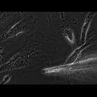
Primary bone marrow macrophages derived from mouse plated on a coverslip and imaged live. Phase contrast microscopy allows us to see the dense f-actin rich leading edge, ruffles, mitochondria, the nu...
CIL:38975
NCBI Organism Classification
Homo sapiens
Biological Process
cell adhesion to artificial surface
Cellular Component
cell surface
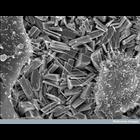
Scanning electron micrograph of two human bone-forming cells (osteoblasts) crawling over crystals of the ceramic material,. monetite (CaHPO4). Monetite crystals are electrochemically deposited onto ti...
CIL:42801
NCBI Organism Classification
none specified
Biological Process
none specified
Cellular Component
cell surface
Colorized scanning electron micrograph of a cell cultured lung cancer cell. This image is part of an image group, CIL 42801-42803, showing several colorized scanning electron micrographs of cell cult...
1
2
3
4
Next »
Results per page:
10
20
50
100