Alternate header for print version
Contributors
Help
Submit
Search
menu
Data sets
Videos
Latest data
Center for Research in Biological Systems
Basic Science Building, Room 1000
University of California, San Diego
9500 Gilman Drive
La Jolla, CA 92093-0608, USA
Voice
: (858) 534-0276
Fax
: (858) 534-7497
Email
: dorloff@ncmir.ucsd.edu
Search Results for
embryonic morphogenesis
(38 results)
CIL:11821
NCBI Organism Classification
Danio rerio
Biological Process
anatomical structure morphogenesis
Cellular Component
nucleus
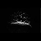
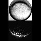
Yolk syncytial layer nuclei undergo epiboly and converge dorsally during late gastrulation. Video made available by Mark Cooper through Zebrafish - The Living Laboratory.
CIL:41659
NCBI Organism Classification
Danio rerio
Biological Process
embryo development
Cellular Component
cell surface
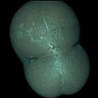
Video of zebrafish embryo development from a 2-cell egg to fish larva. Images were collected using confocal microscopy. Honorable Mention, 2011 Olympus BioScapes Digital Imaging Competition®.
CIL:41911
NCBI Organism Classification
Arabidopsis thaliana
Biological Process
embryonic morphogenesis
Cellular Component
cell wall
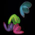
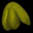
Arabidopsis thaliana (thale cress) embryos imaged with confocal microscopy. Honorable Mention, 2009 Olympus BioScapes Digital Imaging Competition®.
CIL:11805
NCBI Organism Classification
Danio rerio
Biological Process
anatomical structure morphogenesis
Cellular Component
nucleus
Morphogenesis of the ventral yolk syncytial layer (YSL) from sphere stage to the end of gastrulation. The last round of nuclear division occurs before marginal band contraction and epiboly begins. As ...
CIL:11803
NCBI Organism Classification
Danio rerio
Biological Process
gastrulation
Cellular Component
nucleus
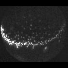
Timelapse of dorsal yolk syncytial layer (YSL) nuclei and blastoderm morphogenesis from sphere stage (late blastula) to 1 somite stage. Top panel shows Nomarski movie of blastoderm morphogenesis inclu...
CIL:41820
NCBI Organism Classification
Arabidopsis thaliana
Biological Process
embryonic morphogenesis
Cellular Component
cell wall
3D stereoscopic projection of a z-stack of confocal images of an Arabidopsis thaliana plant embryo that was fixed, cleared and stained. Red/Green 3D lenses required for stereoscopic view. Honorable Me...
CIL:39788
NCBI Organism Classification
Lytechinus pictus
Biological Process
embryonic morphogenesis
Cellular Component
cell surface
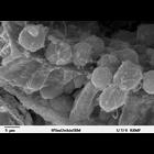
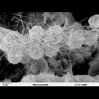
Scanning electron microscope image of Strongylocentrotus drobachiensus embryo at the primary mesenchyme blastula stage. Embryo was split open to reveal the outer epithelial layer and the blastocoel ca...
CIL:39790
NCBI Organism Classification
Lytechinus pictus
Biological Process
embryonic morphogenesis
Cellular Component
cell surface
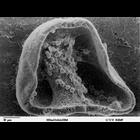
Scanning electron microscope image of Strongylocentrotus drobachiensus [sea urchin] gastrula. This is a higher magnification image of CIL39765. The embryo was split open to reveal a nice cross-section...
CIL:39789
NCBI Organism Classification
Lytechinus pictus
Biological Process
syncytial ring formation
Cellular Component
cell surface
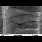
Scanning electron microscope image of Strongylocentrotus drobachiensus [sea urchin] embryo at the late gastrula stage. This is a high magnification image of CIL 39785. Embryo was cut to reveal blastoc...
CIL:39784
NCBI Organism Classification
Lytechinus pictus
Biological Process
cell migration
Cellular Component
cell surface
Scanning electron microscope image of Strongylocentrotus drobachiensus [sea urchin] at the gastrula stage. Embryo was cut in half to reveal blastocoel cavity, containing blastocoel matrix material, pr...
1
2
3
4
Next »
Results per page:
10
20
50
100