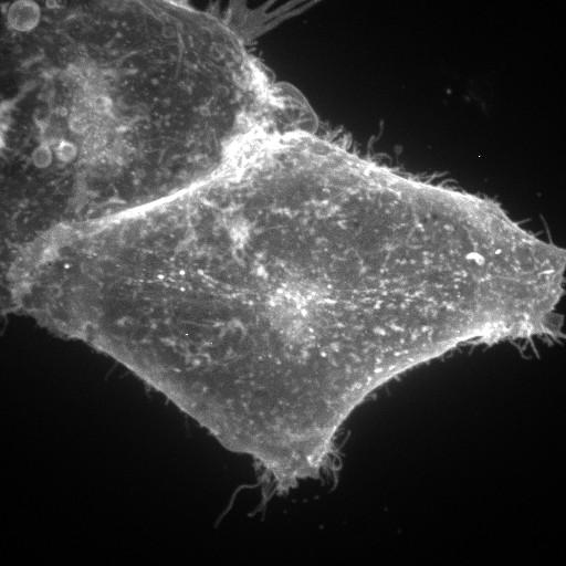
Hela cells transiently transfected with EGFP-GL-GPI show the organization of the membrane thru the distribution of GPI anchored protein. This image is the maximum z projection image that accompanies the related z stack. The images were collected on a Zeiss spinning disk confocal microscope using a 63 X 1.4 NA oil immersion objective. 56 z slices at 0.27 micron intervals were collected with a 450 ms exposure using a 488 ht.
| Spatial Axis | Image Size | Pixel Size |
|---|---|---|
| X | 512px | 0.11µm |
| Y | 512px | 0.11µm |
| Z | —— | 0.27µm |