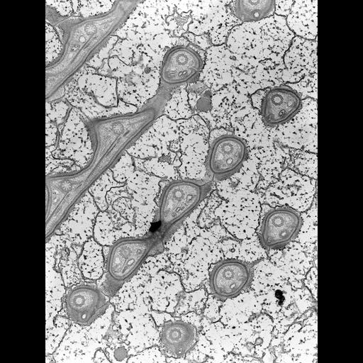
High resolution image of Nassula sp. showing rows of cross-sectioned basal bodies at the cell surface. TEM taken on 3/4/69 by R. Allen with Philips 300 operating at 60kV. Neg. 16,000X. The raw film was scanned with a Nikon Coolscan 9000ED. This image is suitable for quantitative analysis. Standard glutaraldehyde fixation followed by osmium tetroxide, dehydrated in alcohol and embedded in an epoxy resin. Microtome sections prepared at approximately 75nm thickness. Additional information available at (http://www5.pbrc.hawaii.edu/allen/).
| Spatial Axis | Image Size | Pixel Size |
|---|---|---|
| X | 4674px | 0.94nm |
| Y | 6234px | 0.94nm |