Alternate header for print version


Licensing
Attribution Only: This image is licensed under a Creative Commons Attribution License. View License Deed | View Legal Code
tweet  
*CIL – Cell Image Library accession number. Please use this to reference an image.

CIL:57535*  Cite 
Description

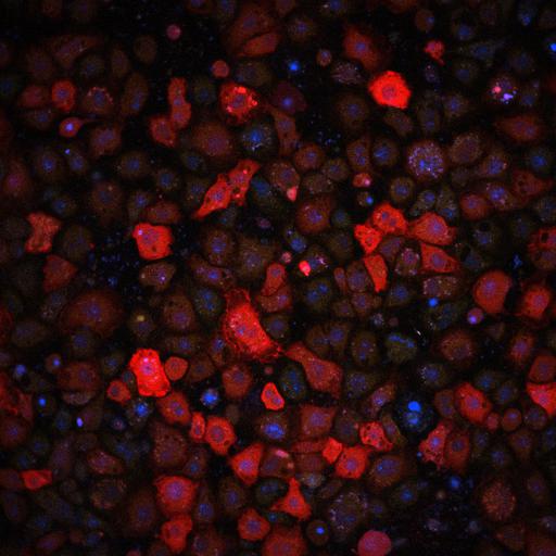
Live-cell imaging of NF-κB immune response in Drosophila S2* cells 405 minutes after stimulus, including nuclear NF-κB and transcription of Dpt gene.

Technical Details

Cells were adhered to glass-bottom wells coated with concanavalin A (a lectin protein), and stimulated with 100 µg/mL DAP-type peptidoglycan (PGN).

Microscopy Setup
Imaging was performed on a Nikon A1R point-scanning confocal microscope equipped with high-sensitivity GaAsP PMTs and a 60× oil immersion objective (NA = 1.4). Acquisition was controlled via Nikon Elements software.

Imaging Parameters
Time-lapse acquisition:

  • 405 minutes following PGN addition
  • Every 30 seconds for 2 hours
  • Image dimensions: 294.6 × 294.6 µm, Resolution: 3.4756 pixels per µm
Biological Sources
NCBI Organism Classification
Drosophila melanogaster
Cell Type
S2* Cells
Cellular Component
cell with labeled nuclei
Relish NF-κB proteins
RhoBAST RNA aptamer expression downstream of immune-responsive enhancer
Biological Context
Biological Process
Peptidoglycan stimulation of IMD innate immune pathway
NF-κB translocation
RNA transcrption
Attribution
Names
Noshin Nawar
Emma Rits
Lianne B. Cohen
Zeba Wunderlich
Link
Article
Github
Citation
Digital Object Identifier (DOI)
doi:10.7295/W9CIL57535
Archival Resource Key (ARK)
ark:/b7295/w9cil57535
Grouping This image is part of a group.
Imaging
Image Mode
point scanning confocal microscopy
time lapse microscopy
Visualization Methods
Channel 1: Hoechst 33258-labeled nuclei
Channel 2: Halotag-labeled Relish (NF-κB) proteins, stained with JF₆₅₀
Channel 3: RhoBAST RNA aptamers under immune-responsive enhancers, labeled with SpyRHO₅₅₅