The biophysical properties of sensory neurons are influenced by their morphometric and morphological features, whose precise measurements require high-quality volume electron microscopy (EM). However, systematic surveys of these nanoscale characteristics for identified neurons are scarce. Taking advantage of the CryoChem method, which permits high-quality ultrastructural preservation of cryofixed and genetically labeled samples for volume EM, we acquired serial block-face scanning electron microscopy (SBEM) images of antennal tissues in which select ORNs expressed the APEX2 EM marker. These SEBEM datasets allow a systematic morphometric and morphological analysis of identified olfactory receptor neurons in Drosophila melanogaster.
Drosophila antennae were prepared as described in Tsang et al. After cryofixation by high-pressure freezing and freeze-substitution in a cocktail containing uranyl acetate and glutaraldehyde, cryofixed antennae were rehydrated gradually at ice temperature. Rehydrated samples were then subjected to DAB labeling reaction and stained using a high-contrast en bloc staining protocol. Next, the samples were dehydrated for resin infiltration and embedded in Durcupan epoxy, followed by imaging with X-ray microscopy and then serial block-face scanning electron microscopy.
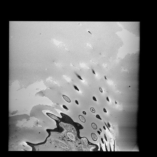
The antenna was imaged using a Zeiss Merlin scanning electron microscope equipped with a Gatan 3View2XP and OnPoint backscatter detector. Images were acquired at 2.5 kV accelerating voltage with a 30 μm condenser aperture and 1.0 μsec dwell time; Z step size was 40 nm; pixel size was 4.11 nm; raster size was 14k x 14k; Z dimension was 1559 sections, and local gas injection was set to 85%. Volume was aligned using cross correlation, segmented, and visualized using IMOD.