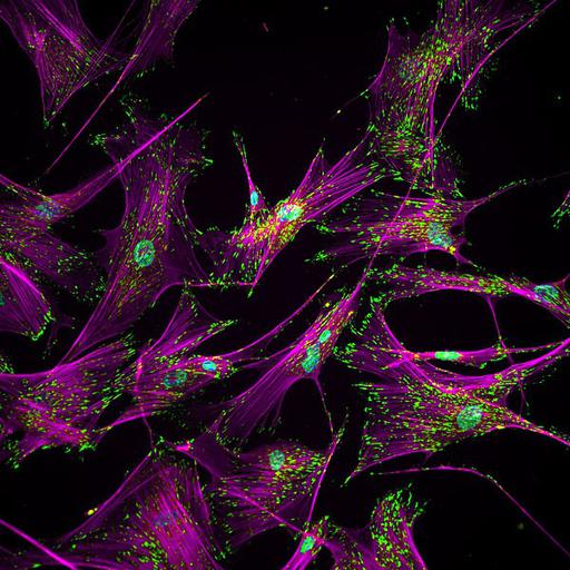
Immunofluorescence image of human IMR90 lung fibroblasts stained for vinculin (green) and filamentous actin (magenta). Nuclei are stained blue. This image of untreated fibroblasts comes from a study of the changes in adhesion that accompany treatment to induce stem cells and can be used in hiPSC purification. See also Singh et al. 2013 Nat Meth 10:438-444. The image appeared in the May 2013 issue of the NIGMS Biomedical Beat which features noteworthy NIGMS-supported research.
IMR90 cells in culture were treated with a cytoskeleton stabilizing buffer, extracted briefly with 0.5% Triton X-100, fixed with 4% paraformaldehyde, and immunostained stained for vinculin using a secondary antibody conjugated with Alexa 488 (green). The actin cytoskeleton was stained with phalloidin conjugated with Alexa 555 (magenta). Nuclei were counterstained with DAPI (blue). Preparations were observed using a Nikon E400 fluorescence microscope with 20x PLANAPO objective lens, and recorded with a SPOT CCD camera. For further details, see A Singh et al. 2013 Adhesion strength-based, label-free isolation of human pluripotent stem cells. Nat Meth 10:438-444 and associated Supplementary Material.
| Spatial Axis | Image Size | Pixel Size |
|---|---|---|
| X | 600px | —— |
| Y | 600px | —— |