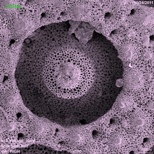
Scanning electron micrograph of the calcium skeleton of coral polyp. The sample was cleaned with bleach solution, dried and carbon coated prior to imaging. The image was collected using a secondary electron detector on the scanning electron microscope, which was operated at 10kV. The original magnification of the image was 100X.
| Spatial Axis | Image Size | Pixel Size |
|---|---|---|
| X | 1920px | —— |
| Y | 1920px | —— |