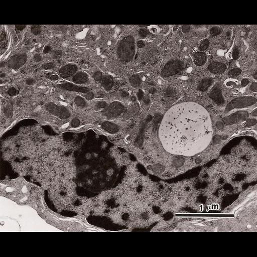
A transmission electron micrograph of a Leydig cell isolated from guinea pig testes. This is a higher magnification image of CIL: 41111 showing nuclear peripheral hererochromatin, highly organized smooth endoplasmic reticulum, and prominent mitochondria.
This image was originally published as Figure 5 in Kukucka, Mark A. and Hara P. Misra. Andrologia 26(4):217-24 (1994), which has been published in final form at: http://onlinelibrary.wiley.com/doi/10.1111/j.1439-0272.1994.tb00791.x/abstract;jsessionid=2082D75B22193F4707988B0AFCE34D3C.d03t04
| Spatial Axis | Image Size | Pixel Size |
|---|---|---|
| X | 965px | —— |
| Y | 780px | —— |