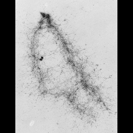
High voltage (1MeV) transmission electron microscopy image of an isolated metaphase chromatid pair from a mouse A9 fibroblast, showing the fiber-like structure.
Cells in metaphase were exposed to 75 mM KCl, spread on water, picked up on grids, stained with uranyl acetate and critical point dried. See also: H. Ris 1981 Stereoscopic electron microscopy of chromosomes. Meth Cell Biol 22:77-96 H. Ris 1978 Preparation of chromatin and chromosomes for electron microscopy. Meth Cell Biol 18:220-246.
| Spatial Axis | Image Size | Pixel Size |
|---|---|---|
| X | 4025px | 2.5nm |
| Y | 5244px | 2.5nm |