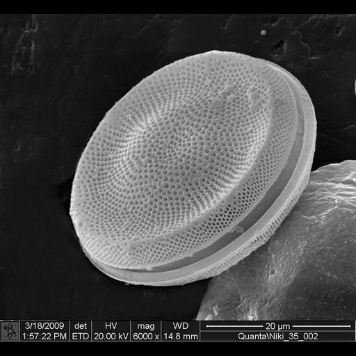
Scanning electron micrograph of a diatom showing the two silica based frustule (cell walls). Image also illustrates the regular pattern on the frustule.
Image collected on a FEI instrument: Quanta Family using Magnification: 6000x, Horizontal Field Width: 20 μm, Voltage: 20 kV, Detector: ETD, and Working Distance: 14.8 mm.
| Spatial Axis | Image Size | Pixel Size |
|---|---|---|
| X | 670px | —— |
| Y | 617px | —— |