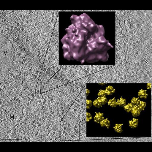
A 5 nm tomographic slice from a vitreous section of a Saccharomyces cerevisiae cell. (M) is a Mitochondrion and (V) a vacuole. Scale bar, 100 nm. Upper Panel: A high-resolution density map of the averaged 80S ribosome constructed using the ribosomes seen in the background image. Lower Panel: A select area from the 80S ribosome Macromolecular Atlas displaying a putative polyribosome cluster
Image collected on a FEI Titan Krios.
| Spatial Axis | Image Size | Pixel Size |
|---|---|---|
| X | 670px | —— |
| Y | 583px | —— |