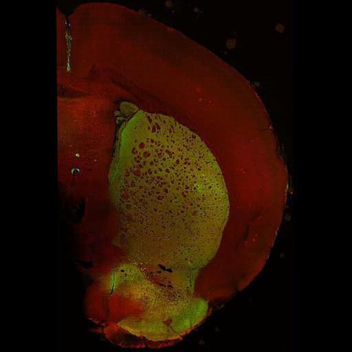
Large scale brain mosaic showing the distribution of the dopamine receptor-associated phosphoprotein DARRP-32 (green) and the vesicular monamine transporter VMAT-2 (red) in a hemisection through the anterior neostriatum of a wild type mouse brain. This image was generated by tiling 45 by 65 (X, Y axes) frames of maximum projection images in the Z-axis. This image has been downsampled from the raw data image which can be accessed using the link provided to the Cell Centered Database.
C57BL6/129SvJ wild type mice from Duke University were perfused with aldehyde fixatives (4% paraformaldehyde + 0.1% gluteraldehyde), sectioned on Vibratome (thickness, 80 µm), and rinsed in phosphate buffered saline (PBS,3 x 10min) and incubated in blocking buffer (PBS, 3% normal donkey serum, 1% fish skin gelatin, 0.3% Triton X-100, 1% bovine serum albumin). Tissue sections incubated on shaker overnight, 4° C in primary antibodies diluted in blocking buffer as follows: anti-VMAT-2; Host = guinea pig; 1:500 (Oncogene, catalog # 503-01-50); anti-DARPP-32; Host = mouse; 1:500 (BD Transduction Laboratories, catalog #611520). Following 3 x 10' rinses, tissue was incubated for 2hr, RT, on a shaker in secondary antibodies diluted at 1:100 in working buffer (donkey-anti-mouse AF488 [Molecular Probes, Cat #A21202] and donkey-anti-guinea pig RRX [Jackson Immunoresearch Laboratories, Inc]. Following rinses in PBS, sections were mounted on slides with gelvatol. Individual images were gathered using a BioRad RTS 2000 Multiphoton with a Nikon PlanApo objective (N.A. 1.45).
| Spatial Axis | Image Size | Pixel Size |
|---|---|---|
| X | 512px | —— |
| Y | 512px | —— |