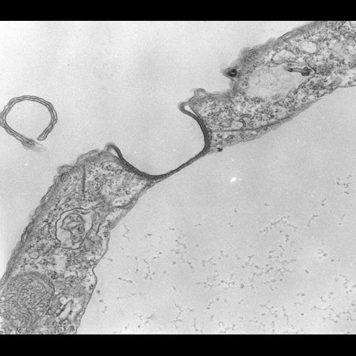
The contractile vacuole pore is coated on its cytosolic side by helically wound microtubules. Other microtubules abut this helix and extend out to the surface of the contractile vacuole where they are attached to the CV membrane and hold the contractile vacuole in place. The plasma membrane and CV membrane are separated only by a narrow gap at the bottom of the pore. TEM taken on 2/11/71 by R. Allen with Hitachi HU11A operating at 60kV. Neg. 19,500X. The raw negative was scanned with an Epson Perfection V750 Pro and this high resolution image is best used for quantitative analysis. Additional information available at (http://www5.pbrc.hawaii.edu/allen/).
Standard glutaraldehyde fixation followed by osmium tetroxide, dehydrated in alcohol and embedded in an epoxy resin. Microtome sections prepared at approximately 75nm thickness. Additional information available at (http://www5.pbrc.hawaii.edu/allen/).
| Spatial Axis | Image Size | Pixel Size |
|---|---|---|
| X | 4654px | 0.77nm |
| Y | 4104px | 0.77nm |