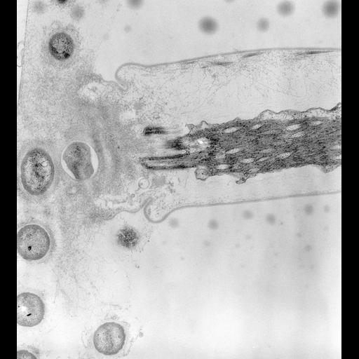
At its most distal extremity the stalk ends in a clump of sheath-like fibers with embedded bacteria. The spasmoneme itself seems to end in a tight association with its surrounding plasma membrane. This is possibly the only point where the myonemal fibers meet the plasma membrane without an intervening alveolus. However there may be microtubules or modified basal bodies linking the spasmonemal fibers to the plasma membrane. The outer surface of the plasma membrane shows striations (at high magnification) next to the holdfast. TEM taken on 2/16/72 by R. Allen with Hitachi HU11A operating at 75kV. Neg. 19,500X. Published and adapted with permission from J. Cell Biol. 56:559-579, 1973. The raw negative was scanned with an Epson Perfection V750 Pro and this high resolution image is best used for quantitative analysis. Additional information available at (http://www5.pbrc.hawaii.edu/allen/).
Standard glutaraldehyde fixation followed by osmium tetroxide, dehydrated in alcohol and embedded in an epoxy resin. Microtome sections prepared at approximately 75nm thickness. Additional information available at (http://www5.pbrc.hawaii.edu/allen/).
| Spatial Axis | Image Size | Pixel Size |
|---|---|---|
| X | 3360px | 1nm |
| Y | 3711px | 1nm |