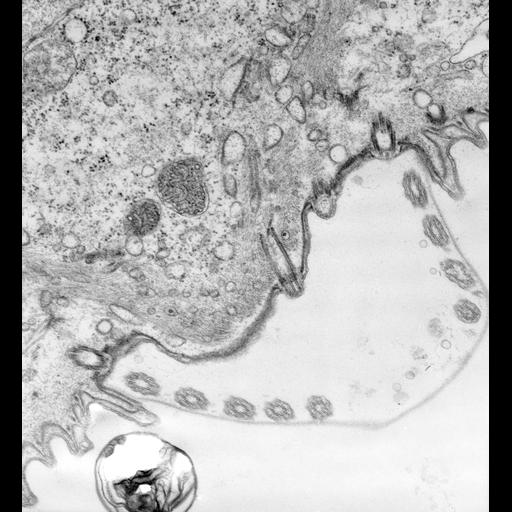
The scopula, stalk-forming region, is outlined by a circle of basal bodies that produce short modified cilia. Other basal bodies lie within this circle. The sheath is secreted outside the circle of cilia. Fibers from linkage complexes enter the proximal ends of these basal bodies. T EM taken on 2/24/72 by R. Allen with Hitachi HU11A operating at 75kV. Neg. 19,500X. The raw negative was scanned with an Epson Perfection V750 Pro and this high resolution image is best used for quantitative analysis. Additional information available at (http://www5.pbrc.hawaii.edu/allen/).
Standard glutaraldehyde fixation followed by osmium tetroxide, dehydrated in alcohol and embedded in an epoxy resin. Microtome sections prepared at approximately 75nm thickness. Additional information available at (http://www5.pbrc.hawaii.edu/allen/).
| Spatial Axis | Image Size | Pixel Size |
|---|---|---|
| X | 4559px | 0.77nm |
| Y | 5004px | 0.77nm |