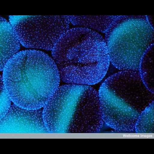
Fluorescent micrograph of stage V-VI Xenopus laevis oocytes surrounded by thousands of follicle cells, as visualized by Hoechst staining.
B0008308 Xenopus laevis oocytes. Wellcome Images available under the following creative commons usage http://creativecommons.org/licenses/by-nc-nd/2.0/uk/
| Spatial Axis | Image Size | Pixel Size |
|---|---|---|
| X | 719px | —— |
| Y | 576px | —— |