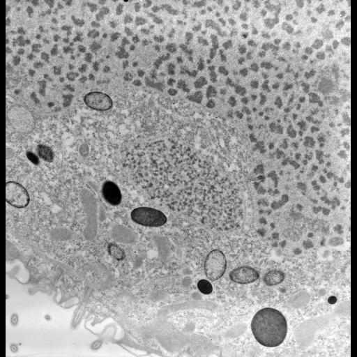
The macronucleus, one micronucleus, and the cytoplasm of Conchophthirus. The negative magnification is 6,800X. The raw film was scanned with an Epson Perfection V750 Pro. This image is best used for quantitative analysis. For greater detail see:Antipa, G. A. 1971. Structural differentiation in the somatic cortex of a ciliated protozoan, Conchophthirus curtus Engelmann, 1862. Protistologica 7:47l-501.
Standard glutaraldehyde fixation followed by osmium tetroxide, dehydrated in alcohol and embedded in an epoxy resin. Microtome sections prepared at approximately 65nm thickness.
| Spatial Axis | Image Size | Pixel Size |
|---|---|---|
| X | 3865px | 1.5nm |
| Y | 4000px | 1.5nm |