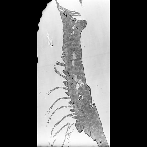
The transition from 3 to 2 rows of cilia can be seen in this view of the AZM. TEM taken on 7/24/67 by R. Allen with Philips 200 operating at 60kV. Neg. 1,370X. Bar = 5µm. Standard glutaraldehyde fixation followed by osmium tetroxide, dehydrated in alcohol, and embedded in an epoxy resin. Microtome sections prepared at approximately 75nm thickness. The raw film was scanned with a Nikon Coolscan 9000ED. This image is suitable for quantitative analysis. Additional information is available at (http://www5.pbrc.hawaii.edu/allen/).
| Spatial Axis | Image Size | Pixel Size |
|---|---|---|
| X | 1972px | 3.7nm |
| Y | 4000px | 3.7nm |