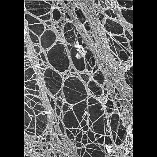
Figure 438 from Chapter 16 (Cytoplasmic matrix and cytoskeleton) of 'The Cell, 2nd Ed.' by Don W. Fawcett M.D. Two bundles of actin-rich stress fibers are apparent in this high magnification micrograph from a 3T3 cell, treated with Triton, washed, quick frozen with liquid helium, deep etched, and rotary shadowed. Image by John Heuser. A PDF copy of the accompanying chapter is available on the ASCB’s BioEDUCATE website.
| Spatial Axis | Image Size | Pixel Size |
|---|---|---|
| X | 886px | —— |
| Y | 1279px | —— |