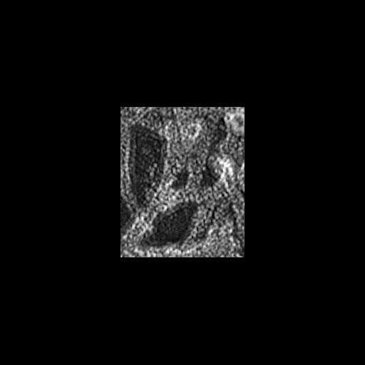
Multiple branching of actin filaments in lamellipodia of vertebrate fibroblasts. This image shows a local enlargement of the leading edge shown in overview in CIL 24788. Image corresponds to Figure 1m from J Cell Biol. 1999 May 31;145(5):1009-26. Figures 1i-o correspond to CIL 24888-24893.
Procedures for detergent extraction, immunostaining, S1 decoration, light, and EM were described previously (Svitkina et al., 1995, 1996, 1997;Verkhovsky et al., 1995; Svitkina and Borisy, 1998).
| Spatial Axis | Image Size | Pixel Size |
|---|---|---|
| X | 174px | —— |
| Y | 211px | —— |