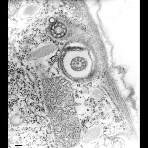
An image of the cortex of Tetrahymena showing a longitudinal microtubular ribbon sandwiched between the alveoli and the epiplasm in the crest of the pellicular ridge. A parasomal sac, the distal end of basal body, the proximal end of cilium, a mucocyst, and mitochondrion are all visible in this micrograph. TEM taken on 1/12/71 by M. Aihara with Hitachi HU11A operating at 75kV. Neg. 17,000X. Bar = 0.2µm. The negative was printed to paper and the image was scanned to Photoshop. This digitized image is available for qualitative analysis. An unprocessed, high resolution version of this image (CIL:34727) is in the library and available for quantitative analysis. Additional information available at (http://www5.pbrc.hawaii.edu/allen/).
Standard glutaraldehyde fixation followed by osmium tetroxide, dehydrated in alcohol and embedded in an epoxy resin. Microtome sections prepared at approximately 75nm thickness. Additional information available at (http://www5.pbrc.hawaii.edu/allen/).
| Spatial Axis | Image Size | Pixel Size |
|---|---|---|
| X | 2982px | —— |
| Y | 3352px | —— |