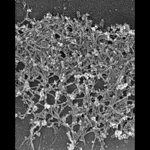
Localization of Xenopus ADF/cofilin (XAC) to posterior regions of depolymerization-resistant actin brush. Electron micrograph of lamellipodia of Xenopus keratocytes after latrunculin a treatment ( 0.25 μM for 10 min) and subsequent staining with XAC antibody. Image correspond to Figure 10 c from J Cell Biol. 1999 May 31;145(5):1009-26.
Procedures for detergent extraction, immunostaining, S1 decoration, light, and EM were described previously (Svitkina et al., 1995, 1996, 1997;Verkhovsky et al., 1995; Svitkina and Borisy, 1998).
| Spatial Axis | Image Size | Pixel Size |
|---|---|---|
| X | 2082px | —— |
| Y | 2609px | —— |