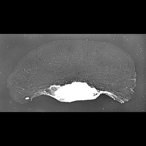
Localization of XAC (Xenopus ADF/cofilin) in Xenopus keratocytes done with immuno-EM. A higher mag view of a localized region of the cell is shown in CIL 24808. Image corresponds to Figure 8g from J Cell Biol. 1999 May 31;145(5):1009-26. A fluorescence image showing XAC and phalloidin labelin is available as CIL 24807.
Procedures for detergent extraction, immunostaining, S1 decoration, light, and EM were described previously (Svitkina et al., 1995, 1996, 1997;Verkhovsky et al., 1995; Svitkina and Borisy, 1998).
| Spatial Axis | Image Size | Pixel Size |
|---|---|---|
| X | 2820px | —— |
| Y | 1512px | —— |