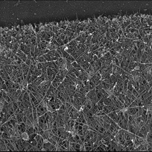
Multiple branching of actin filaments in lamellipodia of Xenopus keratocytes. This image shows an overview of the leading edge, and CIL 24787 shows enlargements of local regions of this platinum replica. CIL 24788 shows a similar overview for the lamellipodia of a vertebrate fibroblasts. Image corresponds to Figure 1a from J Cell Biol. 1999 May 31;145(5):1009-26.
Procedures for detergent extraction, immunostaining, S1 decoration, light, and EM were described previously (Svitkina et al., 1995, 1996, 1997;Verkhovsky et al., 1995; Svitkina and Borisy, 1998).
| Spatial Axis | Image Size | Pixel Size |
|---|---|---|
| X | 3286px | —— |
| Y | 3268px | —— |