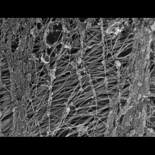
Preferential distribution of plectin in peripheral regions. REF-52 cells were treated with nocodazole to depolymerize MTs and induce collapse of most IFs to the cell center. Electron microscopy of perinuclear cell regions after gelsolin treatment and immunogold (10 nm) labeling for plectin. shows perinuclear intermediate filament bundles are smooth and contain little plectin. Electron microscopy of cytoskeletons was performed as described (Svitkina et al., 1995). Briefly, cells on coverslips were lysed as for light microscopy, treated, with recombinant gelsolin NHz-terminal domain, fixed with glutaraldehyde, tannic acid and uranyl acetate, critical point dried, and coated with platinum and carbon. Image corresponds to Fig 4d from J Cell Biol. 1996 Nov;135(4):991-1007
| Spatial Axis | Image Size | Pixel Size |
|---|---|---|
| X | 4146px | —— |
| Y | 3183px | —— |