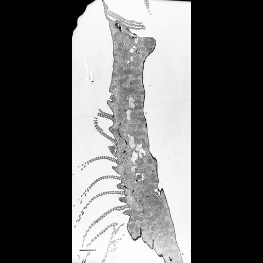
The transition from 3 to 2 rows of cilia can be seen in this view of the AZM. TEM taken on 7/24/67 by R. Allen with Philips 200 operating at 60kV. Neg. 1,370X. Bar = 5µm. Standard glutaraldehyde fixation followed by osmium tetroxide, dehydrated in alcohol, and embedded in an epoxy resin. Microtome sections prepared at approximately 75nm thickness. The negative was scanned and processed in Photoshop. This image is best used for qualitative analysis. A high resolution image is available in the library at (CIL:38890) for qualitative analysis. Additional information is available at (http://www5.pbrc.hawaii.edu/allen/).
| Spatial Axis | Image Size | Pixel Size |
|---|---|---|
| X | 1387px | —— |
| Y | 2055px | —— |