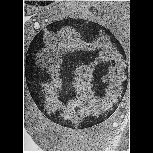
Transmission electron micrograph illustrating the accumulation of dense heterochromation that accompanies terminal differentiation in may cells. Shown here is a section through a polychromatophilic erythroblast from guinea pig bone marrow. The exclusion of heterochromatin from nuclear pore regions is clearly seen. Figure 115 from Chapter 4 (Nucleus) of 'The Cell, 2nd Ed.' by Don W. Fawcett M.D. A PDF copy of the corresponding chapter is available on the ASCB's BioEDUCATE website.
| Spatial Axis | Image Size | Pixel Size |
|---|---|---|
| X | 891px | —— |
| Y | 1272px | —— |