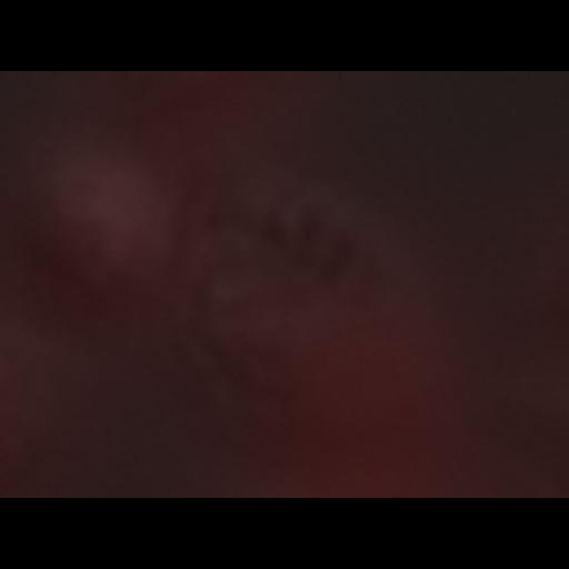
Macrophage cell line cell infected with Leishmania mexicana. Five amastigotes can be seen within a single large parasitophorous vacuole in the macrophage. Raw 264.7 cells (a leukaemic mouse macrophage line) were infected with amastigote form Leishmania mexicana (WHO strain MNYC/BZ/62/M379) at an MOI of 10 and maintained in RPMI for 4 days. Cells were fixed with 2% paraformaldehyde, and DNA was labelled with propidium iodide following incubation with 50 ug/ml RNaseA for 30 min at room temperature. Z stack of 93 images with a z spacing of 0.194 um captured with a 100x/1.4 NA objective lens on a DM5500 B epifluorescence microscope (Leica) with an Orca cooled CCD camera (Hamamatsu). Note there is a phase channel image that is not currently displayed, but can be turned on in the full viewer mode.
| Spatial Axis | Image Size | Pixel Size |
|---|---|---|
| X | 640px | 0.0641µm |
| Y | 480px | 0.0641µm |
| Z | 93px | 0.194nm |
| Channel | Wavelength | |
|---|---|---|
| 1 | dic, fluorescencenm |