Alternate header for print version
Contributors
Help
Submit
Search
menu
Data sets
Videos
Latest data
Center for Research in Biological Systems
Basic Science Building, Room 1000
University of California, San Diego
9500 Gilman Drive
La Jolla, CA 92093-0608, USA
Voice
: (858) 534-0276
Fax
: (858) 534-7497
Email
: dorloff@ncmir.ucsd.edu
Search Results for
yolk
(79 results)
CIL:11811
NCBI Organism Classification
Danio rerio
Biological Process
cell migration
Cellular Component
nucleus
Short timelapse movie of prechordal plate, hypoblast, and yolk syncytial layer (YSL) nuclear movements during 80-90% epiboly. The prechordal plate and notochord can be seen as a tighter clump of cells...
CIL:11805
NCBI Organism Classification
Danio rerio
Biological Process
anatomical structure morphogenesis
Cellular Component
nucleus
Morphogenesis of the ventral yolk syncytial layer (YSL) from sphere stage to the end of gastrulation. The last round of nuclear division occurs before marginal band contraction and epiboly begins. As ...
CIL:37411
NCBI Organism Classification
Danio rerio
Biological Process
anatomical structure morphogenesis
Cellular Component
cytoplasm
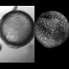
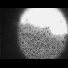
Zebrafish embryo at 80% epiboly. The initial frame shows the junction of the enveloping layer with the yolk cell. The next frame is at a focal plane deeper within the embryo. The blastoderm margin is ...
CIL:11825
NCBI Organism Classification
Danio rerio
Biological Process
anterior/posterior axis specification
Cellular Component
none specified
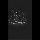
Visualization of yolk syncytial layer (YSL) nuclei in living zebrafish embryos. Sytox Green, a membrane-impermiant vital nuclear stain, was injected into the yold cell of late blastula (sphere stage)...
CIL:37405
NCBI Organism Classification
Danio rerio
Biological Process
anatomical structure morphogenesis
Cellular Component
nucleus
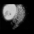
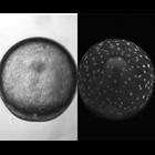
Dome-stage Zebrafish embryo. The animal pole is at the bottom of the image. The yolk syncytial layer shows two tiers of nuclei. As the sequence begins, the yolk syncytial layer narrows. The view then ...
CIL:11809
NCBI Organism Classification
Danio rerio
Biological Process
cell migration
Cellular Component
nucleus
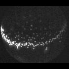
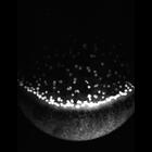
Nomarski/confocal projection shows animal pole of view of head anlage and underlying movements of yolk syncytial layer (YSL) nuclei (bright spots). Nuclei extend and flow through animal pole region to...
CIL:11812
NCBI Organism Classification
Danio rerio
Biological Process
anatomical structure morphogenesis
Cellular Component
nucleus
Timelapse shows yolk syncytial layer (YSL) nuclear migration on the ventral side during gastrulation. Divergence of YSL nuclei away from ventral midline can be seen. As epiboly ends and blastopore clo...
CIL:11801
NCBI Organism Classification
Danio rerio
Biological Process
anatomical structure morphogenesis
Cellular Component
nucleus
Timelapse of dorsal yolk syncytial layer (YSL) nuclear movements during zebrafish gastrulation. A marginal band of YSL nuclei undergo epiboly toward the vegetal pole while internal-YSL (I-YSL) nuclei ...
CIL:35991
NCBI Organism Classification
Rana pipiens
Biological Process
embryo development
Cellular Component
yolk
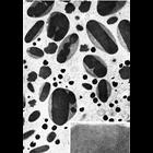
Figure 370 from Chapter 15 (Cytoplasmic Inclusions) of 'The Cell, 2nd Ed.' by Don W. Fawcett M.D. Ectoderm cell from a blastula of Rana pipiens. This cell shows a number of yolk platelets, lipid dro...
CIL:39024
NCBI Organism Classification
Danio rerio
Biological Process
gene expression
Cellular Component
PARD3
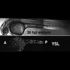
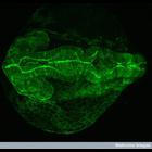
Confocal micrograph of a transgenic zebrafish embryo at 24 hours post-fertilization, showing expression of the fusion protein PARD3_GFP. PARD proteins, which were first identified in C. elegans, are e...
« Previous
1
2
3
4
5
6
7
8
Next »
Results per page:
10
20
50
100