Alternate header for print version
Contributors
Help
Submit
Search
menu
Data sets
Videos
Latest data
Center for Research in Biological Systems
Basic Science Building, Room 1000
University of California, San Diego
9500 Gilman Drive
La Jolla, CA 92093-0608, USA
Voice
: (858) 534-0276
Fax
: (858) 534-7497
Email
: dorloff@ncmir.ucsd.edu
Search Results for
ventral mesoderm
(106 results)
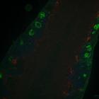
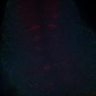
CIL:54672
NCBI Organism Classification
Drosophila melanogaster
Biological Process
Glial-Glial Tiling
Cellular Component
Astrocyte membranes
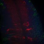
Confocal image of the CNS of a D. melanogaster in the third instar larval stage after Spz3 KD, 44uM from the ventral surface, depicting astrocytes (red), cortex glia (green) and neuronal nuclei (blue)...
CIL:11809
NCBI Organism Classification
Danio rerio
Biological Process
cell migration
Cellular Component
nucleus
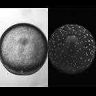
Nomarski/confocal projection shows animal pole of view of head anlage and underlying movements of yolk syncytial layer (YSL) nuclei (bright spots). Nuclei extend and flow through animal pole region to...
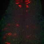
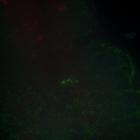
CIL:54655
NCBI Organism Classification
Drosophila melanogaster
Biological Process
Glial-Glial Tiling
Cellular Component
Astrocyte membranes
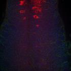
Confocal image of the CNS of a control D. melanogaster in the third instar larval stage, 22uM from the ventral surface, depicting astrocytes (red), cortex glia (green) and neuronal nuclei (blue). Our ...
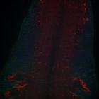
CIL:54656
NCBI Organism Classification
Drosophila melanogaster
Biological Process
Glial-Glial Tiling
Cellular Component
Astrocyte membranes
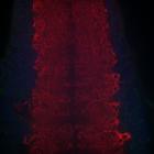
Confocal image of the CNS of a control D. melanogaster in the third instar larval stage, 56uM from the ventral surface, depicting astrocytes (red), cortex glia (green) and neuronal nuclei (blue). Our ...
CIL:54688
NCBI Organism Classification
Drosophila melanogaster
Biological Process
Glial-Glial Tiling
Cellular Component
Astrocyte membranes
Confocal image of the CNS of a D. melanogaster in the third instar larval stage after Spz3 KD, 60uM from the ventral surface, depicting astrocytes (red), cortex glia (green) and neuronal nuclei (blue)...
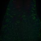
CIL:54658
NCBI Organism Classification
Drosophila melanogaster
Biological Process
Glial-Glial Tiling
Cellular Component
Astrocyte membranes
Confocal image of the CNS of a control D. melanogaster in the third instar larval stage, 36uM from the ventral surface, depicting astrocytes (red), cortex glia (green) and neuronal nuclei (blue). Our ...
CIL:54659
NCBI Organism Classification
Drosophila melanogaster
Biological Process
Glial-Glial Tiling
Cellular Component
Astrocyte membranes
Confocal image of the CNS of a control D. melanogaster in the third instar larval stage, 16uM from the ventral surface, depicting astrocytes (red), cortex glia (green) and neuronal nuclei (blue). Our ...
CIL:54689
NCBI Organism Classification
Drosophila melanogaster
Biological Process
Glial-Glial Tiling
Cellular Component
Astrocyte membranes
Confocal image of the CNS of a D. melanogaster in the third instar larval stage after Spz3 KD, 16uM from the ventral surface, depicting astrocytes (red), cortex glia (green) and neuronal nuclei (blue)...
CIL:54650
NCBI Organism Classification
Drosophila melanogaster
Biological Process
Glial-Glial Tiling
Cellular Component
Astrocyte membranes
Confocal image of the CNS of a control D. melanogaster in the third instar larval stage, 32uM from the ventral surface, depicting astrocytes (red), cortex glia (green) and neuronal nuclei (blue). Our ...
CIL:54648
NCBI Organism Classification
Drosophila melanogaster
Biological Process
Glial-Glial Tiling
Cellular Component
Astrocyte membranes
Confocal image of the CNS of a control D. melanogaster in the third instar larval stage, 16uM from the ventral surface, depicting astrocytes (red), cortex glia (green) and neuronal nuclei (blue). Our ...
« Previous
1
2
3
4
5
6
7
8
9
...
11
Next »
Results per page:
10
20
50
100