Alternate header for print version
Contributors
Help
Submit
Search
menu
Data sets
Videos
Latest data
Center for Research in Biological Systems
Basic Science Building, Room 1000
University of California, San Diego
9500 Gilman Drive
La Jolla, CA 92093-0608, USA
Voice
: (858) 534-0276
Fax
: (858) 534-7497
Email
: dorloff@ncmir.ucsd.edu
Search Results for
retinal outer plexiform layer
(1809 results)
CIL:34538
NCBI Organism Classification
Homo sapiens
Biological Process
cell motility
Cellular Component
none specified
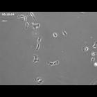
Movie showing motility and morphology of cultured hTERT-RPE1 cells (telomerase immortalized human retinal pigment epithelium) arrested in G1 using 0.08 uM nocodazole (Uetake and Sluder 2010, Curr Biol...
CIL:34848
NCBI Organism Classification
Homo sapiens
Biological Process
mitosis
Cellular Component
none specified
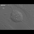
Time lapse movie of cell growth and division in cultured hTERT-RPE1 cells (telomerase immortalized human retinal pigment epithelium) using DIC optics. This movie and the stack of images used to prepar...
CIL:10105
NCBI Organism Classification
Homo sapiens
Biological Process
none specified
Cellular Component
clathrin coat of coated pit
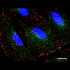
Human retinal pigmented epithelial (RPE) cells labeled for clathrin-coated pits (green), focal adhesions (red) and nuclei (blue). RPE cells stabily expressing 'clathrin light chain a' tagged with EGFP...
CIL:40386
NCBI Organism Classification
Psidium guajava
Biological Process
plant stem organization
Cellular Component
mucilage
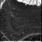
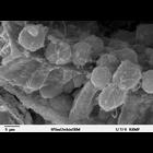
Scanning electron microscope image of cross-section through a Psidium guajava stem. The image shows the thin outer epidermal layer with a thick layer of cortex beneath. The vascular area consists of ...
CCDB:7750
Species
mouse
Organ
eye
Cell type
retinal ganglion cell
System
central nervous system
Structure
cell body
Large scale mosaic imaging with retrograde labeling of retinal ganglion cells
CCDB:8752
Species
mouse
Organ
eye
Cell type
retina rod cell
System
central nervous system
Structure
mitochondrion
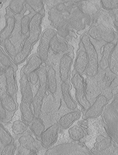
Cone and rod mitochondria: electron tomography
CIL:50521
NCBI Organism Classification
Mydas
Biological Process
none specified
Cellular Component
none specified
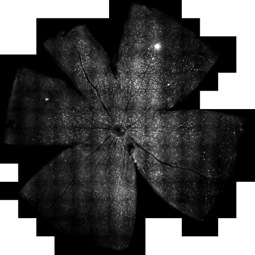
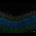
Retinal cross section of C. mydas juvenile from dorsal region with opsins double immunostained with a fluorophore tagged secondary; 1:500 diluted CERN-906 cone opsin (green), 1:500 diluted MAB5356 rho...
CCDB:54
Species
mouse
Organ
eye
Cell type
photoreceptor/cone
System
central nervous system
Structure
mitochondrion
Cone and rod mitochondria: electron tomography
CIL:39788
NCBI Organism Classification
Lytechinus pictus
Biological Process
embryonic morphogenesis
Cellular Component
cell surface
Scanning electron microscope image of Strongylocentrotus drobachiensus embryo at the primary mesenchyme blastula stage. Embryo was split open to reveal the outer epithelial layer and the blastocoel ca...
CIL:39744
NCBI Organism Classification
Mus musculus
Biological Process
photoreception
Cellular Component
nucleus
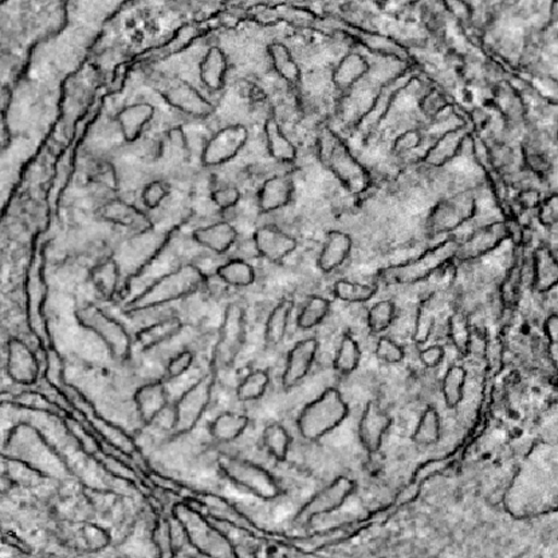
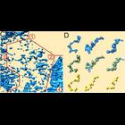
Surface rendered models of chromatin organization from tomogram derived from a TEM image of epon section of a freeze-substituted mouse retina. (C) shows the region boxed in CIL:39743 where 4 regions o...
« Previous
1
...
4
5
6
7
8
9
10
11
...
181
Next »
Results per page:
10
20
50
100