Alternate header for print version
Contributors
Help
Submit
Search
menu
Data sets
Videos
Latest data
Center for Research in Biological Systems
Basic Science Building, Room 1000
University of California, San Diego
9500 Gilman Drive
La Jolla, CA 92093-0608, USA
Voice
: (858) 534-0276
Fax
: (858) 534-7497
Email
: dorloff@ncmir.ucsd.edu
Search Results for
retinal outer plexiform layer
(1809 results)
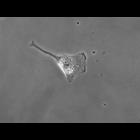
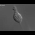
CIL:35151
NCBI Organism Classification
Homo sapiens
Biological Process
mitosis
Cellular Component
none specified
Time lapse series of cell growth and division in cultured hTERT-RPE1 cells (telomerase immortalized human retinal pigment epithelium) using differential interference contrast optics. This movie is gro...
CIL:52244
NCBI Organism Classification
Mus musculus
Biological Process
Retinal aging
Cellular Component
Zonula occludens-1 (ZO-1)
Merged confocal image of mouse (C57BL/6J, postnatal day 330) retinal pigment epithelium (RPE) flatmount. The image is stained for ZO-1 (Green, tight junctions between cells) and nucleus (Red, Propidiu...
CIL:52234
NCBI Organism Classification
Mus musculus
Biological Process
Retinal aging
Cellular Component
Zonula occludens-1 (ZO-1)
Merged confocal image of mouse (C57BL/6J, postnatal day 30) retinal pigment epithelium (RPE) flatmount. The image is stained for ZO-1 (Green, tight junctions between cells) and nucleus (Red, Propidium...
CIL:52239
NCBI Organism Classification
Mus musculus
Biological Process
Retinal aging
Cellular Component
Zonula occludens-1 (ZO-1)
Merged confocal image of mouse (C57BL/6J, postnatal day 60) retinal pigment epithelium (RPE) flatmount. The image is stained for ZO-1 (Green, tight junctions between cells) and nucleus (Red, Propidium...
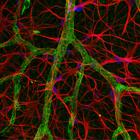
CIL:42509
NCBI Organism Classification
Rattus
Biological Process
retinal aging
Cellular Component
astrocyte projection
Blood vessels and astrocytes in aging rat retina, confocal imaging, 40x. Blood vessels are shown in blue; astrocytes (supportive cells of the nervous system) are mostly in red. As organisms age, ...
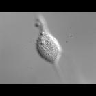
CIL:25693
NCBI Organism Classification
Homo sapiens
Biological Process
mitosis
Cellular Component
none specified
Time series of cell growth and division in cultured hTERT-RPE1 cells (telomerase immortalized human retinal pigment epithelium) using phase contrast optics. A movie created with this series is grouped...
CIL:25694
NCBI Organism Classification
Homo sapiens
Biological Process
mitosis
Cellular Component
none specified
Time lapse series of cell growth and division in cultured hTERT-RPE1 cells (telomerase immortalized human retinal pigment epithelium) using phase contrast optics. A movie created from this time series...
CIL:25707
NCBI Organism Classification
Homo sapiens
Biological Process
mitosis
Cellular Component
none specified
Time lapse series of cell growth and division in cultured hTERT-RPE1 cells (telomerase immortalized human retinal pigment epithelium) using differential interference contrast optics. This time series ...
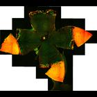
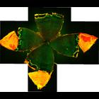
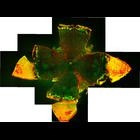
CIL:39008
NCBI Organism Classification
none specified
Biological Process
dendrite morphogenesis
Cellular Component
dendrite
This fluorescent micrograph shows a microelectrode penetrating the cell body of a neurone in the retina of a living eye. The electrode is injecting a fluorescent dye (lucifer yellow) into the cell, fi...
CIL:47050
NCBI Organism Classification
Mus musculus
Biological Process
none specified
Cellular Component
cell body
The purpose of this study was to investigate the progression of changes in retinal ganglion cells and optic nerve glia in neurofibromatosis-1 (NF1) genetically-engineered mice with optic glioma. RGC w...
« Previous
1
...
3
4
5
6
7
8
9
10
...
181
Next »
Results per page:
10
20
50
100


")