Alternate header for print version
Contributors
Help
Submit
Search
menu
Data sets
Videos
Latest data
Center for Research in Biological Systems
Basic Science Building, Room 1000
University of California, San Diego
9500 Gilman Drive
La Jolla, CA 92093-0608, USA
Voice
: (858) 534-0276
Fax
: (858) 534-7497
Email
: dorloff@ncmir.ucsd.edu
Search Results for
retinal outer plexiform layer
(1809 results)
CIL:40380
NCBI Organism Classification
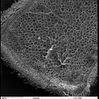
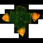
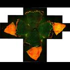
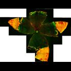
Solenostemon scutellarioides
Biological Process
plant stem organization
Cellular Component
cortex
Scanning electron microscope image of cross-section through a Solenostemon scutellarioidesi (coleus) stem. The image shows the outer epidermal layer, cortex, vascular bundles in a ring, and a central ...
CIL:39745
NCBI Organism Classification
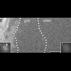
Mus musculus
Biological Process
photoreception
Cellular Component
nucleus
TEM image of epon section of a freeze-substituted mouse retina illustrating the unusual distribution of heterochromatin in rod receptor nuclei where the compact heterochromatin is centrally located. W...
CIL:38803
NCBI Organism Classification
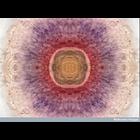
Danio rerio
Biological Process
gene expression
Cellular Component
none specified
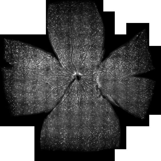
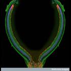
This photomicrograph shows the retina from the eye of a three-day-old zebrafish. The retina is viewed here from the front, as if the viewer is looking directly into the eye of the fish. This image is...
CIL:52249
NCBI Organism Classification
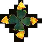
Mus musculus
Biological Process
Retinal aging
Cellular Component
Zonula occludens-1 (ZO-1)
Merged confocal image of mouse (C57BL/6J, postnatal day 720) retinal pigment epithelium (RPE) flatmount. The image is stained for ZO-1 (Green, tight junctions between cells) and nucleus (Red, Propidiu...
CIL:52246
NCBI Organism Classification
Mus musculus
Biological Process
Retinal aging
Cellular Component
Zonula occludens-1 (ZO-1)
Merged confocal image of mouse (C57BL/6J, postnatal day 720) retinal pigment epithelium (RPE) flatmount. The image is stained for ZO-1 (Green, tight junctions between cells) and nucleus (Red, Propidiu...
CIL:52247
NCBI Organism Classification
Mus musculus
Biological Process
Retinal aging
Cellular Component
Zonula occludens-1 (ZO-1)
Merged confocal image of mouse (C57BL/6J, postnatal day 720) retinal pigment epithelium (RPE) flatmount. The image is stained for ZO-1 (Green, tight junctions between cells) and nucleus (Red, Propidiu...
CIL:52238
NCBI Organism Classification
Mus musculus
Biological Process
Retinal aging
Cellular Component
Zonula occludens-1 (ZO-1)
Merged confocal image of mouse (C57BL/6J, postnatal day 60) retinal pigment epithelium (RPE) flatmount. The image is stained for ZO-1 (Green, tight junctions between cells) and nucleus (Red, Propidium...
CCDB:7732
Species
mouse
Organ
eye
Cell type
retinal ganglion cell
System
central nervous system
Structure
cell body
Large scale mosaic imaging with retrograde labeling of retinal ganglion cells
CIL:38802
NCBI Organism Classification
Mus musculus
Biological Process
neural retina development
Cellular Component
nucleus
This confocal micrograph shows the detailed structure of the retina from a one-month-old mouse. The retina is the photoreceptive organ of the eye. It is composed of layers of neuronal cells that captu...
CIL:10136
NCBI Organism Classification
Homo sapiens
Biological Process
clathrin coated pit dynamics
Cellular Component
clathrin coat of coated pit
Visualization of clathrin coated pit dynamics. Total internal reflection fluorescencec microscopy (TIRF-FM) was performed on live human retinal pigment epithelial (RPE) cells stably expressing 'clathr...
« Previous
1
2
3
4
5
6
7
8
9
...
181
Next »
Results per page:
10
20
50
100


")