Alternate header for print version
Contributors
Help
Submit
Search
menu
Data sets
Videos
Latest data
Center for Research in Biological Systems
Basic Science Building, Room 1000
University of California, San Diego
9500 Gilman Drive
La Jolla, CA 92093-0608, USA
Voice
: (858) 534-0276
Fax
: (858) 534-7497
Email
: dorloff@ncmir.ucsd.edu
Search Results for
embryonic structure
(1824 results)
CIL:11822
NCBI Organism Classification
Danio rerio
Biological Process
anatomical structure morphogenesis
Cellular Component
nucleus
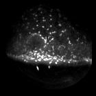
Yolk syncytial layer cell nuclei exhibit convergence and extension behaviors during late gastrulation. Video made available by Mark Cooper through Zebrafish - The Living Laboratory.
CIL:7620
NCBI Organism Classification
Rattus rattus
Biological Process
none specified
Cellular Component
zonula adherens
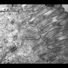
Junctional complex between two cells in the epithelium of the rat intestinal mucosa. The tight junction (also known as zonula occludens) is the structure located nearest to the lumen. The elimination ...
CIL:36038
NCBI Organism Classification
Drosophila melanogaster
Biological Process
meiosis
Cellular Component
synaptonemal complex
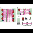
Model of the structure of the synaptonemal complex (SC) in Drosophila females based on EM immunolocalization of SC components C(3)G and C(2)M.
CIL:39090
NCBI Organism Classification
Homo sapiens
Biological Process
proximal tubule morphogenesis
Cellular Component
cell surface
A colorized SEM image of a human proximal tubule, showing the tubular structure and projections extending over the tissue surface.
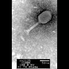
CIL:41130
NCBI Organism Classification
Enterobacteria phage T4
Biological Process
bacteriophage structure
Cellular Component
phage head
Transmission electron microscope image of T4Phage, a bacteriophage that infects Escherichia coli bacteria. This image is part of an image group (CIL:41124-41131).
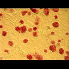
CIL:12630
NCBI Organism Classification
Homo sapiens
Biological Process
pluripotency
Cellular Component
none specified
Human embryonic stem cell colonies that have been stained for alkaline phosphatase (red). Alkaline phosphatase staining is used to show that the colonies are pluripotent. The colonies are growing on a...
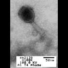
CIL:41125
NCBI Organism Classification
Enterobacteria phage T4
Biological Process
bacteriophage structure
Cellular Component
phage head
Transmission electron microscope image of T4Phage, a bacteriophage that infects Escherichia coli bacteria. This image is part of an image group (CIL:41124-41131).
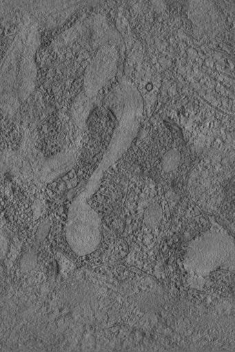
CCDB:8638
Species
rat
Organ
Brain
Cell type
none specified
System
central nervous system
Structure
neuropil
Tomographic imaging of synapses
CIL:39089
NCBI Organism Classification
Plasmodium yoelii nigeriensis
Biological Process
sporozoan zygote development
Cellular Component
cyst wall
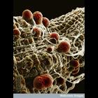
Colorized scanning electron micrograph of malaria (Plasmodium yoelii nigeriensis) oocysts ( thick-walled structure in which sporozoan zygotes develop) developing on the midgut wall of the mosquito Ano...
CIL:40973
NCBI Organism Classification
Drosophila melanogaster
Biological Process
uterus morphogenesis
Cellular Component
none specified
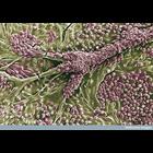
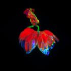
Fluorescent micrograph of a Drosophila melanogaster reproductive system highlighting the muscular and neural structure fruitfly ovaries and uterus. The background staining of the eggs in red is a spe...
« Previous
1
...
6
7
8
9
10
11
12
13
...
183
Next »
Results per page:
10
20
50
100