Alternate header for print version
Contributors
Help
Submit
Search
menu
Data sets
Videos
Latest data
Center for Research in Biological Systems
Basic Science Building, Room 1000
University of California, San Diego
9500 Gilman Drive
La Jolla, CA 92093-0608, USA
Voice
: (858) 534-0276
Fax
: (858) 534-7497
Email
: dorloff@ncmir.ucsd.edu
Search Results for
visualization by chemical attribute
(6422 results)
CIL:13713
NCBI Organism Classification
Homo sapiens
Biological Process
protein phosphorylation
Cellular Component
mitochondrion
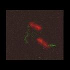
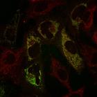
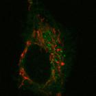
Upon mitochondrial uncoupling, wild-type PINK1-YFP (green) is localized in mitochondria (red) in HeLa cells. HeLa cells expressing WT PINK1-YFP were stained with 10nM MitoTracker Red (red) for 30 min ...
CIL:13715
NCBI Organism Classification
Homo sapiens
Biological Process
protein phosphorylation
Cellular Component
mitochondrion
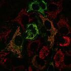
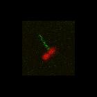
PINK1-YFP R98F (green) is found localized to mitochondria (red) in the absence of the mitochondrial depolarizing agent CCCP (carbonyl cyanide-m-chlorophenyl hydrazone). The R98F mutation in PINK1-YFP ...
CIL:13714
NCBI Organism Classification
Homo sapiens
Biological Process
protein phosphorylation
Cellular Component
mitochondrion
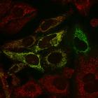
PINK1-YFP R98F (green) is found localized to mitochondria (red) in the presence of the mitochondrial depolarizing agent CCCP (carbonyl cyanide-m-chlorophenyl hydrazone). The R98F mutation in PINK1-YFP...
CIL:7245
NCBI Organism Classification
Homo sapiens
Biological Process
cytokinesis
Cellular Component
microtubule
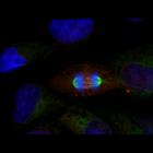
HeLa cell in cytokinesis was fixed and stained for microtubules (green), RacGAP1 (red), and DNA (blue). Cells were fixed with MeOH on ice for 3 min, and stained with a primary antibody against RacGA...
CIL:9070
NCBI Organism Classification
Rattus
Biological Process
mitochondrion organization
Cellular Component
mitochondrion
NRK cells expressing a mitochondria marker, mito-RFP (red), and PSS1-CFP (green), phosphatidylserine synthase 1. A single 6 micron confocal slice was imaged on a Zeiss LSM 510 every 5.9 sec with the...
CIL:7324
NCBI Organism Classification
Escherichia coli
Biological Process
pilus assembly
Cellular Component
pilus
This is one of four related images of F-pili (conjugative pili encoded by the F plasmid of E. coli) dynamics by live-cell imaging. There are 2 movie (.avi) files and 2 files (.tiff) from the movies. ...
CIL:7321
NCBI Organism Classification
Escherichia coli
Biological Process
pilus assembly
Cellular Component
pilus
This is one of four related images of F-pili (conjugative pili encoded by the F plasmid of E. coli) dynamics by live-cell imaging. There are 2 movie (.avi) files and 2 files (.tiff) from the movies. ...
CIL:826
NCBI Organism Classification
Notophthalmus viridescens
Biological Process
ribosomal RNA transcription
Cellular Component
nuclear chromosome
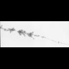
Amphibian oocytes have many copies of rRNA genes that are actively transcribed. Newt oocyte nuclei were dissected, allowed to swell in a low ionic strength high pH buffer, centrifuged through a sucros...
CIL:11636
NCBI Organism Classification
Rattus sp.
Biological Process
epithelial cilium movement
Cellular Component
cilium
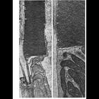
Figures 323 & 324 from Chapter 13 (Cilia and Flagella) of 'The Cell' by Don W. Fawcett M.D. The outer segments of the rods and cones of the vertebrate retina and many photoreceptors of invertebrates b...
CIL:12067
NCBI Organism Classification
Paramecium multimicronucleatum
Biological Process
cortical cytoskeleton organization
Cellular Component
cell cortex
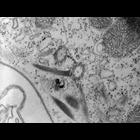
High resolution image of a tangential section of basal body pair (dikinetid) showing a connecting fiber linking the BBs as well as attachments to a kinetodesmal fiber, to postciliary microtubules, an...
« Previous
1
2
3
4
5
6
7
8
9
...
643
Next »
Results per page:
10
20
50
100


")