Alternate header for print version
Contributors
Help
Submit
Search
menu
Data sets
Videos
Latest data
Center for Research in Biological Systems
Basic Science Building, Room 1000
University of California, San Diego
9500 Gilman Drive
La Jolla, CA 92093-0608, USA
Voice
: (858) 534-0276
Fax
: (858) 534-7497
Email
: dorloff@ncmir.ucsd.edu
Search Results for
scanning electron microscopy (SEM)
(381 results)
CIL:11094
NCBI Organism Classification
Phodopus
Biological Process
single fertilization
Cellular Component
microvillus membrane
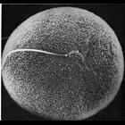
Scanning electron micrograph showing in vitro fertilization of a hamster oocyte. The sperm head, near the center of the field of view, is engulfed by the microvilli on the surface of the oocyte membr...
CIL:11096
NCBI Organism Classification
Phodopus
Biological Process
single fertilization
Cellular Component
microvillus membrane
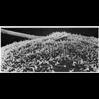
Scanning electron micrograph showing a lateral view of sperm penetration of a hamster oocyte during vitro fertilization. As the membrane of the sperm head fuses with that of the oocyte, the microvill...
CIL:11110
NCBI Organism Classification
Parophrys vetulus
Biological Process
plasma membrane organization
Cellular Component
cell surface
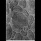
of 'The Cell, 2nd Ed.' by Don W. Fawcett M.D. Scanning electron micrograph of the epidermal surface of the flatfish sole Parophrys detulus shows a labyrinth of surface ridges. Image from Fahrenbach,...
CIL:11108
NCBI Organism Classification
Bufo
Biological Process
extracellular structure organization
Cellular Component
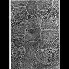
cell surface
Scanning electron micrograph of the lumenal surface of the toad bladder shows the boundaries of four polygonal squamous epithelial cells surrounding a mitochondrial-rich cell bearing short globular mi...
CIL:11113
NCBI Organism Classification
Oncorhynchus kisutch
Biological Process
plasma membrane organization
Cellular Component
cell surface
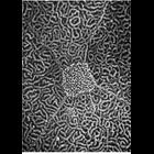
Scanning electron micrograph of the epidermal surface of the Pacific coho salmon. Ridge-like folds of the plasmalemma, called microplicae, display a labyrinth pattern across the surface, and a circum...
CIL:11115
NCBI Organism Classification
Entosphenus tridentatus
Biological Process
plasma membrane organization
Cellular Component
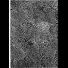
cell surface
Scanning electron micrograph of the epidermal surface of lamprey larvae. A row of microvilli outline the polygonal borders between cells, while short microvilli cover the external surface in a reticu...
CIL:11131
NCBI Organism Classification
none specified
Biological Process
mitosis
Cellular Component
lamellipodium
The final stages of cell division is captured in the scanning electron micrograph. Two daughter cells are still interconnected by slender strands of membrane (white arrow at center of image). Lamell...
CIL:35464
NCBI Organism Classification
Rattus norvegicus
Biological Process
none specified
Cellular Component
none specified
Liver sinusoid of a Brown Rat with fenestrated endothelial cells. Sinusoidal width is about 5 microns. Original magnification is 10,000x . Note the microvilli of hepatocytes in the space of Disse exte...
CIL:35465
NCBI Organism Classification
Rattus norvegicus
Biological Process
none specified
Cellular Component
none specified
Fenestrae of a Brown Rat arranged in sieve plates. Original magnification is 20,000x, a good indication of liver sinudosial endothelial cells (LESCs).
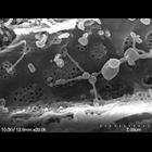
CIL:53296
NCBI Organism Classification
Prokaryote
Biological Process
none specified
Cellular Component
Cell surphace morphology
Control-treated MRSA biofilm
« Previous
1
2
3
4
5
6
7
8
9
...
39
Next »
Results per page:
10
20
50
100