Alternate header for print version
Contributors
Help
Submit
Search
menu
Data sets
Videos
Latest data
Center for Research in Biological Systems
Basic Science Building, Room 1000
University of California, San Diego
9500 Gilman Drive
La Jolla, CA 92093-0608, USA
Voice
: (858) 534-0276
Fax
: (858) 534-7497
Email
: dorloff@ncmir.ucsd.edu
Search Results for
probe for mitochondria
(20 results)
CIL:13707
NCBI Organism Classification
Homo sapiens
Biological Process
mitochondrion degradation
Cellular Component
mitochondrion
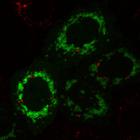
YFP-Parkin (green) localizes to mitochondria (red) when HeLa cells are transfected with control siRNA (siCtrl) and treated with the mitochondrial depolarizing agent CCCP (carbonyl cyanide-m-chlorophen...
CIL:13709
NCBI Organism Classification
Homo sapiens
Biological Process
mitochondrion degradation
Cellular Component
mitochondrion
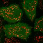
YFP-Parkin (green) does not translocate to mitochondria (red) when HeLa cells are transfected with PARL (presenilin-associated rhomboid-like protein) siRNA. HeLa cells stably expressing YFP-Parkin wer...
CIL:13712
NCBI Organism Classification
Homo sapiens
Biological Process
mitochondrion degradation
Cellular Component
mitochondrion
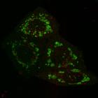
YFP-Parkin (green) localizes to mitochondria (red) when HeLa cells are treated with the mitochondrial depolarizing agent CCCP (carbonyl cyanide-m-chlorophenyl hydrazone). HeLa cells stably expressing ...
CIL:39061
NCBI Organism Classification
Bos taurus
Biological Process
cytoskeleton organization
Cellular Component
mitochondrion
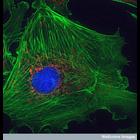
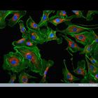
This image shows a bovine pulmonary artery endothelial cell. The pulmonary artery is the large vessel that takes deoxygenated blood cells from the heart to the lungs. These cells have been stained wit...
CIL:39059
NCBI Organism Classification
Bos taurus
Biological Process
cytoskeleton organization
Cellular Component
mitochondrion
This confocal image shows bovine pulmonary artery cells visualized with DAPI to highlight the nucleus (blue), MitoTracker Red CMXRos to stain mitochondria (red dots), and Alexafluor 488 phalloidin to ...
CIL:245
NCBI Organism Classification
Mus musculus
Biological Process
intermediate filament-based process
Cellular Component
mitochondrion
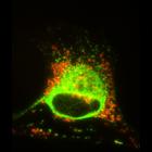
Mitochondrial distribution (visualized with Mitotracker CMXRos) was largely unaffected by partial fragmentation of the intermediate filament cytoskeleton, visualized here with vimentin antibody, after...
CIL:247
NCBI Organism Classification
Mus musculus
Biological Process
intermediate filament-based process
Cellular Component
type III intermediate filament
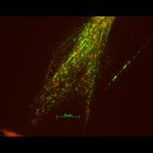
This ghoulish-looking Schwann cell was present in a primary culture of the dorsal root ganglion of mouse that was transfected with a mutant peripherin transgene. The intermediate filament network (vi...
CIL:31203
NCBI Organism Classification
Gallus gallus
Biological Process
axonal transport
Cellular Component
mitochondrion
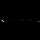
Mitochondrial transport during a growth cone pause. Chicken DRG neurons were grown on coverslips coated with laminin/poly-L-ornithine for 2 d and stained with 0.1 µM MitoTracker Red CMXRos. Images we...
CIL:31202
NCBI Organism Classification
Gallus gallus
Biological Process
axonal transport
Cellular Component
mitochondrion
Mitochondrial transport during axonal elongation. Chicken DRG neurons were grown on coverslips coated with laminin/poly-L-ornithine for 2 d and stained with 0.1 µM MitoTracker red CMXRos. Images were...
CIL:31201
NCBI Organism Classification
Gallus gallus
Biological Process
axonal transport
Cellular Component
mitochondrion
Chicken DRG neurons were grown on coverslips coated with laminin/poly-L-ornithine for 2 d and stained with 0.1 µM MitoTracker red CMXRos. Images were acquired every 2 s. To reduce the size of the vid...
« Previous
1
2
Results per page:
10
20
50
100