Alternate header for print version
Contributors
Help
Submit
Search
menu
Data sets
Videos
Latest data
Center for Research in Biological Systems
Basic Science Building, Room 1000
University of California, San Diego
9500 Gilman Drive
La Jolla, CA 92093-0608, USA
Voice
: (858) 534-0276
Fax
: (858) 534-7497
Email
: dorloff@ncmir.ucsd.edu
Search Results for
primary antibody plus labeled secon...
(2148 results)
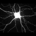
CIL:807
NCBI Organism Classification
Rattus
Biological Process
calcium-dependent cell-cell adhesion
Cellular Component
excitatory synapse
During hippocampal neuron development in vitro, N-cadherin becomes selectively localized to excitatory synapses. This image shows immunolocalization of N-cadherin. Other images in this image group sh...
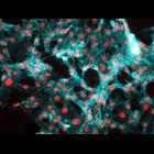
CIL:50647
NCBI Organism Classification
Homo sapiens
Biological Process
none specified
Cellular Component
Vascular endothelial cadherin
Human umbilical vein endothelial cells stained initially for nuclei with DAPI (blue) and for vascular endothelial cadherin (red).
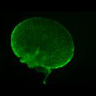
CIL:12627
NCBI Organism Classification
Mus musculus
Biological Process
oocyte maturation
Cellular Component
vasa
Fluorescent image of a mouse ovary labeled with an antibody to vasa (involved in oocyte differentiation and polarity) which is localized in the oocytes within the ovary. The sample was fixed in parafo...
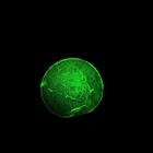
CIL:12632
NCBI Organism Classification
Mus musculus
Biological Process
none specified
Cellular Component
zona pellucida
Mouse zona pellucida. This image shows a zona pellucida surrounding a mouse oocyte. The sample has been labeled with an antibody to ZP3, one of the three glycoproteins in the zona pellucida. The image...
CIL:24797
NCBI Organism Classification
none specified
Biological Process
actin filament-based process
Cellular Component
lamellipodium
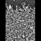
Localization of Arp2/3 complex in the lamellioidia of afibroblast . Staining with p21 antibody and 10 nm gold secondary antibody. CIL 24794 shows a fluorescence microsopy image of Arp2/3 localizatio...
CIL:24806
NCBI Organism Classification
Xenopus laevis
Biological Process
cellular localization
Cellular Component
lamellipodium
Localization of XAC (Xenopus ADF/cofilin) in Xenopus keratocytes. Fluorescence microscopy of a whole cell double stained with XAC antibody (green) and TRITC-phalloidin (red). XAC in lamellipodium is e...
CIL:24785
NCBI Organism Classification
Xenopus laevis
Biological Process
actin filament-based process
Cellular Component
lamellipodium
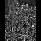
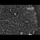
Localization of XAC (Xenopus ADF/cofilin) in Xenopus fibroblasts. Immuno-EM with XAC antibody at high magnification. Low magnification view is available at CIL 24784. Nucleus and surrounding regions...
CIL:24795
NCBI Organism Classification
Xenopus laevis
Biological Process
cellular macromolecule localization
Cellular Component
lamellipodium
Localization of Arp2/3 complex in the lamellioidia of a Xenopus keratocytes. Staining with p21 antibody and 10 nm gold secondary antibody. CIL 24794 shows a fluorescence microsopy image of Arp2/3 lo...
CIL:35618
NCBI Organism Classification
Homo sapiens
Biological Process
phosphorylation of myosin light chain
Cellular Component
actin filament
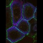
The relationship between the actin cytoskeleton (blue), adherens junctions (green) and phospho-Myosin Light Chain (MLC) (red) after 30 minutes of calcium stimulation.
CIL:36148
NCBI Organism Classification
Drosophila melanogaster
Biological Process
microtubule cytoskeleton organization
Cellular Component
microtubule
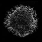
The first image in this multi-image tiff file is a stimulated emission depletion image (STED) of microtubules in a Drosophila S2 cell. The second image is the corresponding diffraction limited image ...
« Previous
1
...
4
5
6
7
8
9
10
11
...
215
Next »
Results per page:
10
20
50
100

")