Alternate header for print version
Contributors
Help
Submit
Search
menu
Data sets
Videos
Latest data
Center for Research in Biological Systems
Basic Science Building, Room 1000
University of California, San Diego
9500 Gilman Drive
La Jolla, CA 92093-0608, USA
Voice
: (858) 534-0276
Fax
: (858) 534-7497
Email
: dorloff@ncmir.ucsd.edu
Search Results for
permeabilized tissue
(2365 results)
CIL:24803
NCBI Organism Classification
Xenopus laevis
Biological Process
cellular localization
Cellular Component
lamellipodium
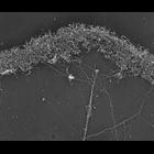
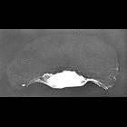
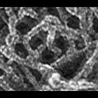
Structural differentiation of actin network in lamellipodium. Electron micrograph of Xenopus fibroblast after unprotected extraction without polyethelene glycol and phalloidin. Actin network at lamel...
CIL:34891
NCBI Organism Classification
none specified
Biological Process
branching of actin filaments
Cellular Component
actin cytoskeleton
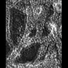
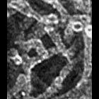
Multiple branching of actin filaments in lamellipodia of vertebrate fibroblasts. This image shows a local enlargement of the leading edge shown in overview in CIL 24788. Image corresponds to Figure 1...
CIL:34893
NCBI Organism Classification
none specified
Biological Process
branching of actin filaments
Cellular Component
actin cytoskeleton
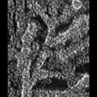
Multiple branching of actin filaments in lamellipodia of vertebrate fibroblasts. This image shows a local enlargement of the leading edge shown in overview in CIL 24788. Image corresponds to Figure 1...
CIL:34901
NCBI Organism Classification
none specified
Biological Process
branching of actin filaments
Cellular Component
actin cytoskeleton
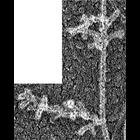
Improved visualization of actin filament branching in lamellipodia. EM of keratocyte or fibroblast lamellipodial actin network after cytochalasin D treatment (0.2 μM for 30 min or 0.5 μM for 10 min)...
CIL:39061
NCBI Organism Classification
Bos taurus
Biological Process
cytoskeleton organization
Cellular Component
mitochondrion
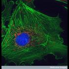
This image shows a bovine pulmonary artery endothelial cell. The pulmonary artery is the large vessel that takes deoxygenated blood cells from the heart to the lungs. These cells have been stained wit...
CIL:42508
NCBI Organism Classification
none specified
Biological Process
cytoskeleton organization
Cellular Component
microtubule cytoskeleton
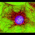
Digital multi-color immuno fluorescence microscopy and phase contrast image of fibroblast cells in culture stained with various antibodies. The Phase contrast (green) image was overlayed over the micr...
CIL:24807
NCBI Organism Classification
Xenopus laevis
Biological Process
cellular localization
Cellular Component
lamellipodium
Localization of XAC (Xenopus ADF/cofilin) in Xenopus keratocytes done with immuno-EM. A higher mag view of a localized region of the cell is shown in CIL 24808. Image corresponds to Figure 8g from J...
CIL:24783
NCBI Organism Classification
Xenopus laevis
Biological Process
cellular localization
Cellular Component
lamellipodium
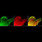
Localization of XAC (Xenopus ADF/cofilin) in Xenopus fibroblasts. Fluorescence microscopy of a cell fragment double stained with XAC antibody (green) and TRITC-phalloidin (red). XAC is distributed t...
CIL:34883
NCBI Organism Classification
Xenopus laevis
Biological Process
branching of actin filaments
Cellular Component
actin cytoskeleton
Multiple branching of actin filaments in lamellipodia of Xenopus keratocytes. This image show an enlargement of a local region from the overview of the leading edge, CIL 24786. Image corresponds to F...
CIL:34887
NCBI Organism Classification
Xenopus laevis
Biological Process
branching of actin filaments
Cellular Component
actin cytoskeleton
Multiple branching of actin filaments in lamellipodia of Xenopus keratocytes. This image show an enlargement of a local region from the overview of the leading edge, CIL 24786. Image corresponds to F...
« Previous
1
2
3
4
5
6
7
8
9
...
237
Next »
Results per page:
10
20
50
100