Alternate header for print version
Contributors
Help
Submit
Search
menu
Data sets
Videos
Latest data
Center for Research in Biological Systems
Basic Science Building, Room 1000
University of California, San Diego
9500 Gilman Drive
La Jolla, CA 92093-0608, USA
Voice
: (858) 534-0276
Fax
: (858) 534-7497
Email
: dorloff@ncmir.ucsd.edu
Search Results for
labeled primary antibody
(159 results)
CIL:7239
NCBI Organism Classification
Homo sapiens
Biological Process
cytokinesis
Cellular Component
microtubule
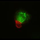
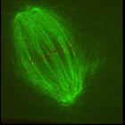
HeLa cells were accumulated in monopolar mitosis using a 12 hr treatment of the kinesin-5 inhibitor S-trityl-l-cysteine and forced into cytokinesis by adding the potent Cdk1 inhibitor purvalanol A for...
CIL:7244
NCBI Organism Classification
Homo sapiens
Biological Process
cytokinesis
Cellular Component
microtubule
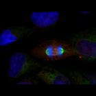
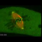
HeLa cells at last stage of cytokinesis were fixed and stained with microtubules (green), PRC1 (red), and KIF4A (blue). PRC1 and KIF4A colocalized on midbody in the center of intracellular bridge and ...
CIL:13090
NCBI Organism Classification
Xenopus [NCBITaxon:262014]
Biological Process
mitotic anaphase
Cellular Component
spindle microtubule
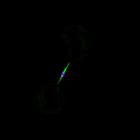
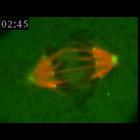
Dual-channel spinning-disk confocal time-lapse movie showing kinetochores (Alexa488 labeled CENP-A antibody) in anaphase (red) relative to poleward microtubule flux. Fluorescent speckle microscopy (FS...
CIL:7245
NCBI Organism Classification
Homo sapiens
Biological Process
cytokinesis
Cellular Component
microtubule
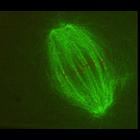
HeLa cell in cytokinesis was fixed and stained for microtubules (green), RacGAP1 (red), and DNA (blue). Cells were fixed with MeOH on ice for 3 min, and stained with a primary antibody against RacGA...
CIL:13089
NCBI Organism Classification
Xenopus [NCBITaxon:262014]
Biological Process
mitotic metaphase
Cellular Component
spindle microtubule
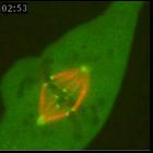
Dual-channel spinning-disk confocal time-lapse movie showing kinetochores (Alexa488 labeled CENP-A antibody) in metaphase (red) relative to poleward microtubule flux. Fluorescent speckle microscopy (F...
CIL:12020
NCBI Organism Classification
Potorous tridactylus
Biological Process
attachment of spindle microtubules to kinetochore
Cellular Component
kinetochore
Mitotic PtK1 cell microinjected with X-rhodamine-labeled tubulin and Alexa 488-labeled CENP-F antibodies to fluorescently label kinetochore fibers (red) and kinetochores/spindle poles (green), respect...
CIL:12019
NCBI Organism Classification
Potorous tridactylus
Biological Process
attachment of spindle microtubules to kinetochore
Cellular Component
kinetochore
Mitotic PtK1 cell microinjected with X-rhodamine-labeled tubulin and Alexa 488-labeled CENP-F antibodies to fluorescently label kinetochore fibers (red) and kinetochores/spindle poles (green), respect...
CIL:8078
NCBI Organism Classification
Potorous tridactylus
Biological Process
attachment of spindle microtubules to kinetochore
Cellular Component
kinetochore
Metaphase PtK1 cell microinjected with X-rhodamine-labeled tubulin and Alexa 488-labeled CENP-F antibodies, to fluorescently label kinetochore fibers (red) and kinetochores/spindle poles (green), resp...
CIL:8079
NCBI Organism Classification
Potorous tridactylus
Biological Process
attachment of spindle microtubules to kinetochore
Cellular Component
kinetochore
The movie starts in metaphase and shows sister kinetochore pairs aligned at the metaphase plate and oscillating between the two spindle poles as their respective kinetochore fibers lengthen and shorte...
« Previous
1
...
8
9
10
11
12
13
14
15
16
Results per page:
10
20
50
100