Alternate header for print version
Contributors
Help
Submit
Search
menu
Data sets
Videos
Latest data
Center for Research in Biological Systems
Basic Science Building, Room 1000
University of California, San Diego
9500 Gilman Drive
La Jolla, CA 92093-0608, USA
Voice
: (858) 534-0276
Fax
: (858) 534-7497
Email
: dorloff@ncmir.ucsd.edu
Search Results for
crosslinking-fixative fixed tissue
(3962 results)
CIL:11403
NCBI Organism Classification
Felis catus
Biological Process
cellular respiration
Cellular Component
mitochondrion
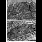
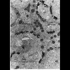
Figures 222 (upper) and 223 (lower) from Chapter 7 (Mitochondria) of 'The Cell, 2nd Ed.' by Don W. Fawcett M.D. Mitochondrial matrix granules are located within the space bounded by the inner mitocho...
CIL:10839
NCBI Organism Classification
Phodopus
Biological Process
autophagy
Cellular Component
lysosome
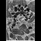
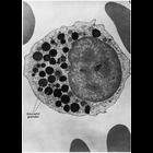
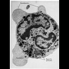
Figure 263 from Chapter 8 (Lysosomes) of 'The Cell, 2nd Ed.' by Don W. Fawcett M.D. Portion of a cell from hamster suprarenal cortex with clustered lysosomes near the Golgi complex. A PDF copy of th...
CIL:11106
NCBI Organism Classification
Rattus
Biological Process
cell projection organization
Cellular Component
brush border
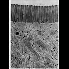
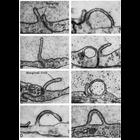
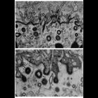
Electron micrograph showing the brush border of intestinal epithelial cells of the rat. The parallel microvilli of the brush border are about 0.1 µm wide and 1.0 µm in length. Image by Jean Paul R...
CIL:11137
NCBI Organism Classification
Mammalia
Biological Process
pinocytosis
Cellular Component
vesicle
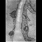
Capillary endothelial cells from mammalian cardiac muscle caught in the act of fluid-phase micropinocytosis. Arrows indicate multiple sites where invaginaton along the membrane suggests vesicles formi...
CIL:11362
NCBI Organism Classification
Mus musculus
Biological Process
post-translational protein modification
Cellular Component
Golgi apparatus
Figure 199 from Chapter 6 (Golgi Apparatus) of 'The Cell, 2nd Ed.' by Don W. Fawcett M.D. The Golgi apparatus in epithelial cells from the mouse epididymis. The convex, or cis, side of the Golgi sho...
CIL:11135
NCBI Organism Classification
Felis catus
Biological Process
pinocytosis
Cellular Component
plasma membrane
Electron micrographs show examples of pinocytosis in endothelial cells of blood vessels in the cat myocardium (panels A,B,C and H), and from the choroid rete of the bowfin fish eye. Both types of cell...
CIL:11150
NCBI Organism Classification
Homo sapiens
Biological Process
pinocytosis
Cellular Component
coated vesicle
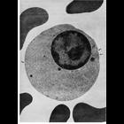
This transmission electron micrograph of a section through a late orthochromatic erythroblast (normoblast) from human bone marrow shows shallow depressions along the membrane surface, indicated by arr...
CIL:10851
NCBI Organism Classification
Homo sapiens
Biological Process
immune system process
Cellular Component
azurophil granule
Figure 276 from Chapter 8 (Lysosomes) of 'The Cell, 2nd Ed.' by Don W. Fawcett M.D. An early myelocyte from human bone marrow with numerous azurophil granules with cytochemical properties of lysosome...
CIL:11164
NCBI Organism Classification
Periplaneta americana
Biological Process
vitellogenesis
Cellular Component
coated vesicle
Uptake of yolk protein into coated vesicles (arrows) at the periphery of oocytes of the cockroach Periplaneta americana. Yolk protein binds to regions of the membrane with a prominent glycocalyx. Beca...
CIL:11123
NCBI Organism Classification
Cavia porcellus
Biological Process
cell motility
Cellular Component
pseudopodium
Pseudopods jut out from this polymorphonuclear leucocyte of the guinea pig to enable amoeboid motility. Organelles are initially excluded as the pseudopod first forms, but they flow in as the pseudopo...
1
2
3
4
5
6
7
8
9
...
397
Next »
Results per page:
10
20
50
100