Alternate header for print version
Contributors
Help
Submit
Search
menu
Data sets
Videos
Latest data
Center for Research in Biological Systems
Basic Science Building, Room 1000
University of California, San Diego
9500 Gilman Drive
La Jolla, CA 92093-0608, USA
Voice
: (858) 534-0276
Fax
: (858) 534-7497
Email
: dorloff@ncmir.ucsd.edu
Search Results for
distribution of a specific protein
(2878 results)
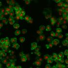
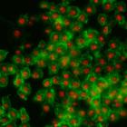
CIL:32137
NCBI Organism Classification
Drosophila melanogaster
Biological Process
cellular localization
Cellular Component
nucleus
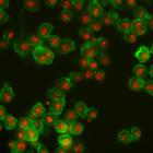
Drosophila melanogaster Kc167 cells were stained for DNA (to label nuclei, red) and actin (a cytoskeletal protein, to show the cell body, green). Each image is a dual channel fluorescent image followe...
CIL:32144
NCBI Organism Classification
Drosophila melanogaster
Biological Process
cellular localization
Cellular Component
nucleus
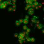
Drosophila melanogaster Kc167 cells were stained for DNA (to label nuclei, red) and actin (a cytoskeletal protein, to show the cell body, green). Each image is a dual channel fluorescent image followe...
CIL:32138
NCBI Organism Classification
Drosophila melanogaster
Biological Process
cellular localization
Cellular Component
nucleus
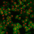
Drosophila melanogaster Kc167 cells were stained for DNA (to label nuclei, red) and actin (a cytoskeletal protein, to show the cell body, green). Each image is a dual channel fluorescent image followe...
CIL:32143
NCBI Organism Classification
Drosophila melanogaster
Biological Process
cellular localization
Cellular Component
nucleus
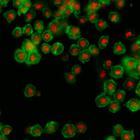
Drosophila melanogaster Kc167 cells were stained for DNA (to label nuclei, red) and actin (a cytoskeletal protein, to show the cell body, green). Each image is a dual channel fluorescent image followe...
CIL:32145
NCBI Organism Classification
Drosophila melanogaster
Biological Process
cellular localization
Cellular Component
nucleus
Drosophila melanogaster Kc167 cells were stained for DNA (to label nuclei, red) and actin (a cytoskeletal protein, to show the cell body, green). Each image is a dual channel fluorescent image followe...
CIL:32131
NCBI Organism Classification
Drosophila melanogaster
Biological Process
cellular localization
Cellular Component
nucleus
Drosophila melanogaster Kc167 cells were stained for DNA (to label nuclei, red) and actin (a cytoskeletal protein, to show the cell body, green). Each image is a dual channel fluorescent image followe...
CIL:10101
NCBI Organism Classification
Homo sapiens
Biological Process
none specified
Cellular Component
epsin
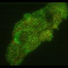
Cultured retinal pigment epithelial cell were stably transfected with mCherry-clathrin light chain and EGFP-epsin. Live-cell time lapse images were collected on a Nikon TiE TIRF with a 100x1.49 NA ob...
CIL:10468
NCBI Organism Classification
Toxoplasma gondii RH
Biological Process
regulation of cell shape
Cellular Component
cortical microtubule
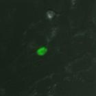
A monolayer of human foreskin fibroblast cells was infected with Toxoplasma gondii expressing EGFP-tagged Tgbeta3-tubulin. Images overlaid with DIC. Images acquired using API DeltaVision on an Olympus...
CIL:10471
NCBI Organism Classification
Toxoplasma gondii RH
Biological Process
regulation of cell shape
Cellular Component
cortical microtubule
A monolayer of human foreskin fibroblast cells was infected with Toxoplasma gondii expressing EGFP-tagged Tgbeta3-tubulin. Images acquired using API DeltaVision on a Olympus IX70 inverted microscope....
CIL:10474
NCBI Organism Classification
Toxoplasma gondii RH
Biological Process
regulation of cell shape
Cellular Component
cortical microtubule
A monolayer of human foreskin fibroblast cells was infected with Toxoplasma gondii expressing EGFP-tagged Tgbeta3-tubulin. Images acquired using API DeltaVision on a Olympus IX70 inverted microscope....
« Previous
1
2
3
4
5
6
7
8
9
...
288
Next »
Results per page:
10
20
50
100
")