Alternate header for print version
Contributors
Help
Submit
Search
menu
Data sets
Videos
Latest data
Center for Research in Biological Systems
Basic Science Building, Room 1000
University of California, San Diego
9500 Gilman Drive
La Jolla, CA 92093-0608, USA
Voice
: (858) 534-0276
Fax
: (858) 534-7497
Email
: dorloff@ncmir.ucsd.edu
Search Results for
differences in deposition of metal ...
(687 results)
CIL:39344
NCBI Organism Classification
Passiflora edulis
Biological Process
pollen wall organization
Cellular Component
pollen wall
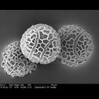
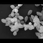
Scanning electron micrograph of passion flower pollen. A related image is CIL:39346.
CIL:40589
NCBI Organism Classification
Malvaceae
Biological Process
none specified
Cellular Component
pollen coat
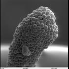
This is a scanning electron micrograph of a Hibiscus pollen grain.
CIL:40655
NCBI Organism Classification
Botryotinia sphaerosperma
Biological Process
none specified
Cellular Component
none specified
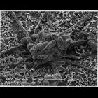
Scanning electron micrograph of Botrytis sp. (a mold) on the skin of a Pinot noir grape. Botrytis sp is grey-mold called nobel rot on wine grapes.
CIL:41311
NCBI Organism Classification
Penta lanceolata
Biological Process
stigma development
Cellular Component
cell surface
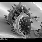
Scanning electron microscope image of Penta lanceolata stigma (receptive surface for pollen).
CIL:12403
NCBI Organism Classification
Leishmania mexicana
Biological Process
amastigote form Leishmania mexicana
Cellular Component
none specified
Leishmania mexicana amastigote forms (WHO strain MNYC/BZ/62/M379). Amastigotes were grown in axenic culture. The cells were fixed with glutaraldehyde, dehydrated in ethanol, critical-point dried and s...
CIL:222
NCBI Organism Classification
none specified
Biological Process
none specified
Cellular Component
plasma membrane
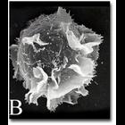
A scanning electron microscope image of an activated mast cell illustrating the convoluted topography of the cell membrane, which is populated with receptors.
CIL:35465
NCBI Organism Classification
Rattus norvegicus
Biological Process
none specified
Cellular Component
none specified
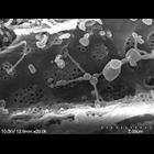
Fenestrae of a Brown Rat arranged in sieve plates. Original magnification is 20,000x, a good indication of liver sinudosial endothelial cells (LESCs).
CIL:38811
NCBI Organism Classification
none specified
Biological Process
none specified
Cellular Component
cell surface
Scanning electron micrograph of red blood cells clearly showing their biconcave disc shape. Human red blood cells are typically 8 microns x 2 microns in size.
CIL:39104
NCBI Organism Classification
none specified
Biological Process
cell-cell adhesion
Cellular Component
cell surface
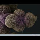
Scanning electron micrograph of pancreatic cancer cells. See additional image at CIL:39076.
CIL:39106
NCBI Organism Classification
none specified
Biological Process
none specified
Cellular Component
cell surface
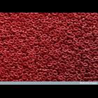
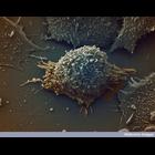
Colorized scanning electron micrograph of a lung cancer cell.
« Previous
1
2
3
4
5
6
7
8
9
...
69
Next »
Results per page:
10
20
50
100