Alternate header for print version
Contributors
Help
Submit
Search
menu
Data sets
Videos
Latest data
Center for Research in Biological Systems
Basic Science Building, Room 1000
University of California, San Diego
9500 Gilman Drive
La Jolla, CA 92093-0608, USA
Voice
: (858) 534-0276
Fax
: (858) 534-7497
Email
: dorloff@ncmir.ucsd.edu
Search Results for
secondary_electron imaging
(49 results)
CIL:35957
NCBI Organism Classification
Leporidae
Biological Process
fertilization
Cellular Component
flagellum
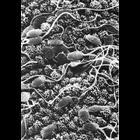
Figure 328 from Chapter 14 (Sperm Flagellum) of 'The Cell, 2nd Ed.' by Don W. Fawcett M.D. Scanning electron micrograph of rabbit spermatozoa on the endometrium of the uterus. Image by David Phillip...
CIL:17893
NCBI Organism Classification
uncultured scuticociliate
Biological Process
cortical cytoskeleton organization
Cellular Component
cilium
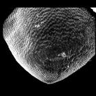
Conchophthirus was deciliated by calcium ion shock followed by shearing through a 22 gauge needle. This revealed the location of the deciliated basal bodies and the cell's surface architecture. In thi...
CIL:39251
NCBI Organism Classification
Didinium nasutum
Biological Process
ciliary cell motility
Cellular Component
oral apparatus
Didinium nasutum showing the two prominent girdles of cilia in full metachronous beat. If you look at higher magnification you will see the occasional short, blunt clavate cilia. These are known to no...
CIL:40904
NCBI Organism Classification
uncultured scuticociliate
Biological Process
cortical cytoskeleton organization
Cellular Component
cilium
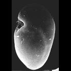
Conchophthirus was deciliated by calcium ion shock followed by shearing through a micropipette. This revealed the location of the deciliated basal bodies and the cell's surface architecture. In this i...
CIL:21994
NCBI Organism Classification
Didinium nasutum
Biological Process
contractile vacuole organization
Cellular Component
contractile vacuole pore
The aboral view of Didinium showing seven contractile vacuole pores for it's single posterior contractile vacuole and metachronous waves of cilia in one of the two characteristic ciliary girdles of Di...
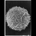
CIL:11105
NCBI Organism Classification
Mus musculus
Biological Process
plasma membrane organization
Cellular Component
cell surface
Scanning electron micrograph of the surface of a peritoneal mast cell from mouse shows undulating folds, and few microvilli. The membrane folds could be misinterpreted for microvilli if analyzed in t...
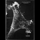
CIL:11129
NCBI Organism Classification
Mus musculus
Biological Process
cell motility
Cellular Component
lamellipodium membrane
Scanning electron micrograph of a 3T3 cell growing in culture. Complexity and diversity of structure across the cell surface is apparent in the this image that s courtesy of Susan Brown. Figure 48 fr...
CIL:11099
NCBI Organism Classification
Mus musculus
Biological Process
immune system process
Cellular Component
microvillus
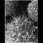
Cells from peritoneal cavity exudate of the mouse, exposed to sodium azide at 22°C for 22 minutes. The macrophage in the field of view has extended long microvilli, showing a rapid response to change...
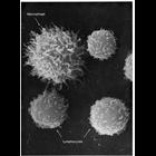
CIL:11102
NCBI Organism Classification
Mus musculus
Biological Process
immune system process
Cellular Component
microvillus
Rapid extension of microvilli on the surface of a peritoneal macrophage, induced by treatment with a metabolic inhibitor, sodium azide, after 20 minutes at 22° C. Microvilli on neighboring lymphocyt...
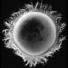
CIL:11094
NCBI Organism Classification
Phodopus
Biological Process
single fertilization
Cellular Component
microvillus membrane
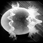
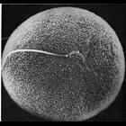
Scanning electron micrograph showing in vitro fertilization of a hamster oocyte. The sperm head, near the center of the field of view, is engulfed by the microvilli on the surface of the oocyte membr...
« Previous
1
2
3
4
5
Next »
Results per page:
10
20
50
100