Alternate header for print version
Contributors
Help
Submit
Search
menu
Data sets
Videos
Latest data
Center for Research in Biological Systems
Basic Science Building, Room 1000
University of California, San Diego
9500 Gilman Drive
La Jolla, CA 92093-0608, USA
Voice
: (858) 534-0276
Fax
: (858) 534-7497
Email
: dorloff@ncmir.ucsd.edu
Search Results for
secondary electron generation
(53 results)
CIL:25356
NCBI Organism Classification
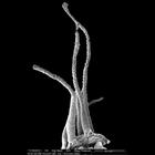
Hydra viridis
Biological Process
none specified
Cellular Component
none specified
Scanning electron micrograph showing tentacles on the head of a Hydra viridis. Epithelial cells located at the base of the tentacle are in a flat, contracted state. Cell distal to the head of the H. v...
CIL:39248
NCBI Organism Classification
Didinium nasutum
Biological Process
phagocytosis
Cellular Component
oral apparatus
Didinium captures Paramecium. Paramecium is almost completely ingested and the proboscis is beginning to reform. This micrograph was taken in 1968 by G. Antipa on a Cambridge Mark IIA operating at 20k...
CIL:40588
NCBI Organism Classification
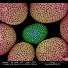
Porifera
Biological Process
structural support
Cellular Component
cell surface
This colorized scanning electron micrograph shows uncoated sponge spicules from a South African sponge. Spicules provide structural support and deter predators. The image was taken with the VcD (back...
CIL:11110
NCBI Organism Classification
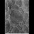
Parophrys vetulus
Biological Process
plasma membrane organization
Cellular Component
cell surface
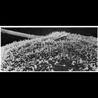
of 'The Cell, 2nd Ed.' by Don W. Fawcett M.D. Scanning electron micrograph of the epidermal surface of the flatfish sole Parophrys detulus shows a labyrinth of surface ridges. Image from Fahrenbach,...
CIL:39249
NCBI Organism Classification
Didinium nasutum
Biological Process
phagocytosis
Cellular Component
oral apparatus
Didinium captures Paramecium. At midingestion and showing metachronous waves of cilia within the two characteristic ciliary girdles of Didinium nasutum. This micrograph was taken in 1968 by G. Antipa ...
CIL:11094
NCBI Organism Classification
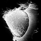
Phodopus
Biological Process
single fertilization
Cellular Component
microvillus membrane
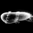
Scanning electron micrograph showing in vitro fertilization of a hamster oocyte. The sperm head, near the center of the field of view, is engulfed by the microvilli on the surface of the oocyte membr...
CIL:11096
NCBI Organism Classification
Phodopus
Biological Process
single fertilization
Cellular Component
microvillus membrane
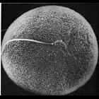
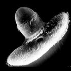
Scanning electron micrograph showing a lateral view of sperm penetration of a hamster oocyte during vitro fertilization. As the membrane of the sperm head fuses with that of the oocyte, the microvill...
CIL:39247
NCBI Organism Classification
Didinium nasutum
Biological Process
phagocytosis
Cellular Component
oral apparatus
Didinium captures Paramecium. After reeling in the prey with cyclosis tugging at the attached toxicysts the proboscis opens to engage the Paramecium. This micrograph was taken in 1968 by G. Antipa on ...
CIL:11099
NCBI Organism Classification
Mus musculus
Biological Process
immune system process
Cellular Component
microvillus
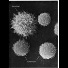
Cells from peritoneal cavity exudate of the mouse, exposed to sodium azide at 22°C for 22 minutes. The macrophage in the field of view has extended long microvilli, showing a rapid response to change...
CIL:11102
NCBI Organism Classification
Mus musculus
Biological Process
immune system process
Cellular Component
microvillus
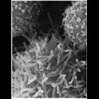
Rapid extension of microvilli on the surface of a peritoneal macrophage, induced by treatment with a metabolic inhibitor, sodium azide, after 20 minutes at 22° C. Microvilli on neighboring lymphocyt...
« Previous
1
2
3
4
5
6
Next »
Results per page:
10
20
50
100